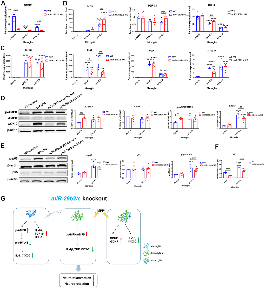
Figure 6.The effects of miR-29b2/c deficiency in LPS-treated primary microglia. (A) qPCR analysis of BDNF (A), IL-10 TGF-β1 and IGF-1 (B), and pro-inflammatory factors IL-1β, IL-6, TNF and COX2 (C) in WT and miR-29b2/c KO primary microglia treated with PBS or LPS for four and eight hours. n=4-6. (D) Western blot analysis of p-AMPK, AMPK and COX-2 protein expression in WT and miR-29b2/c KO primary microglia treated with PBS or LPS for 24 h. β-actin served as a loading control. Quantifications of relative p-AMPK, AMPK and COX-2 protein level and the ratio of p-AMPK to AMPK are shown in the right panel. n=4-6. (E) Western blot analysis of p-p65 and p65 protein expression in WT and miR-29b2/c KO primary microglia treated with PBS or LPS for one hour. Quantifications of relative p-p65 and p65 protein level and their ratio are shown in the right panel. n=4-6. (F) Nitrite concentration in the culture medium of WT and miR-29b2/c KO microglia treated with PBS or LPS for 24 h. n=6. The differences were analyzed by two-way ANOVA followed by LSD multiple comparison tests. *p<0.05, **p < 0.01, ***p<0.001 and ****p<0.0001, vs PBS control. # p < 0.05, ## p < 0.01, ### p<0.001 and ####p<0.0001, vs WT group. (G) Diagram of effects of miR-29b2/c deficiency in Parkinson’s disease.