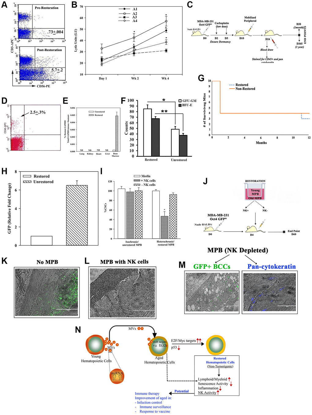
Figure 6.Restorative containing NK cell function. (A) Flow cytometry for CD56+ cells, pre- and post-restoration. (B) Timeline LUs (Supplementary Figure 6A for calculation) in restored cells. (C) Treatment protocol with mice harboring dormant CSCs. (D) Flow cytometry for huCD45+ cells in blood of mice after 1 month of injection with CD3-depleted restored cells. (E) qPCR for huGAPDH at 2 months after injection with CD3-depleted restored cells, mean ± SD (n = 5); ND = not detected. (F) CFU-GM and BFU-E in BM at 1 month post-restoration, mean ± SD, n = 5. *p ≤ 0.05 vs. mice with unrestored cells. (G) Survival curve spanning the study period. (H) At yr 1, qPCR for GFP with cells from femurs. The results presented as fold change in which the lowest value was assigned 1, mean ± SD, 4/group. (I) CSCs, co-cultured with restored MPBs (− or + NK cells) for 24 h. % CSCs (GFP+) were determined by microscopy and flow cytometry, mean ± SD, n = 4. (J) Protocol for NSG with dormant CSCs given restored CD3(−) MPB (−/+ NK cells). (K–M) GFP (surrogate of CSCs) in paraffin section of femurs, −/+ restored MPB: -MPB (K), +MPB (L), MPB without NK cells (M). (N) Summary: Aged hematopoietic cells instructed young cells to produce specific miRNA containing MVs to induce MYC and E2F targets to restore the aged MPBs, leading to increased NK activity. Transplantation of restored cells decreased hallmarks of aging: ↑lymphoid:myeloid, ↓senescence/inflammation. See also Supplementary Figure 6.
Figure 6 — Restoration of aged hematopoietic cells by their young counterparts through instructive microvesicles release | Aging