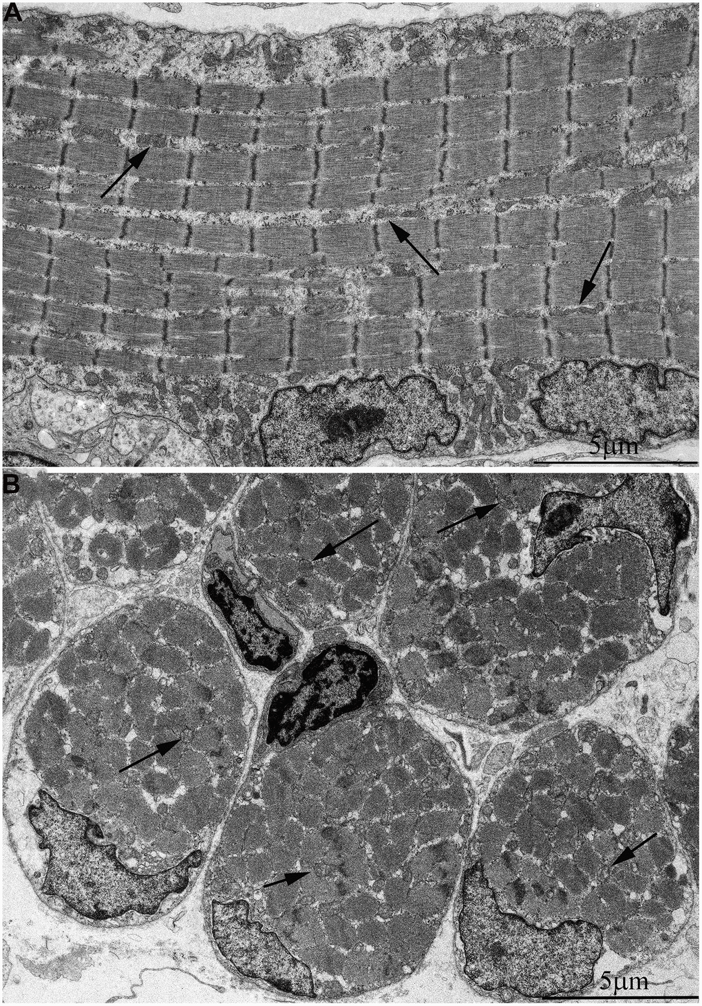
Figure 2.Ultrastructure of mitochondria in skeletal muscle of one-week-old naked mole rat. (A) Longitudinal section. Arrows indicate mitochondria. (B) Cross section. Widely spaced, small mitochondria are observed on the longitudinal and cross section of muscle fiber. Arrows indicate mitochondria.