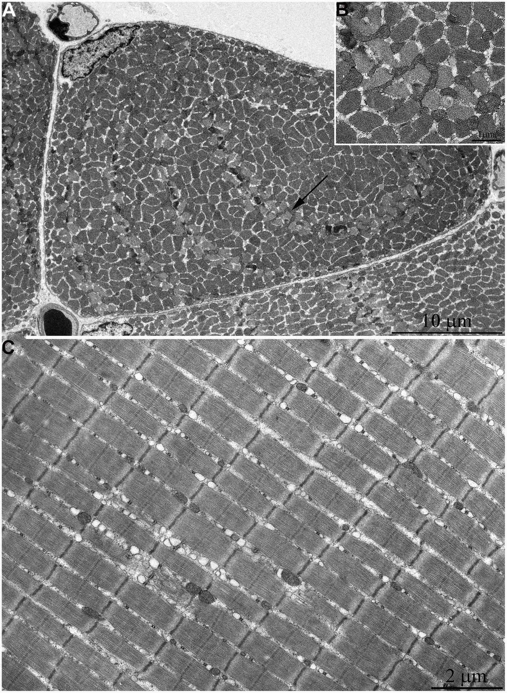
Figure 3.Ultrastructure of mitochondria in skeletal muscle of six-month-old naked mole rat. (A) Cross section of muscle fiber. Small, isolated mitochondria and group of mitochondria, which ultrastructure is demonstrated at higher magnification in (B) is indicated by an arrow. (C) Longitudinal section of muscle fiber. Small, widely spaced mitochondria.