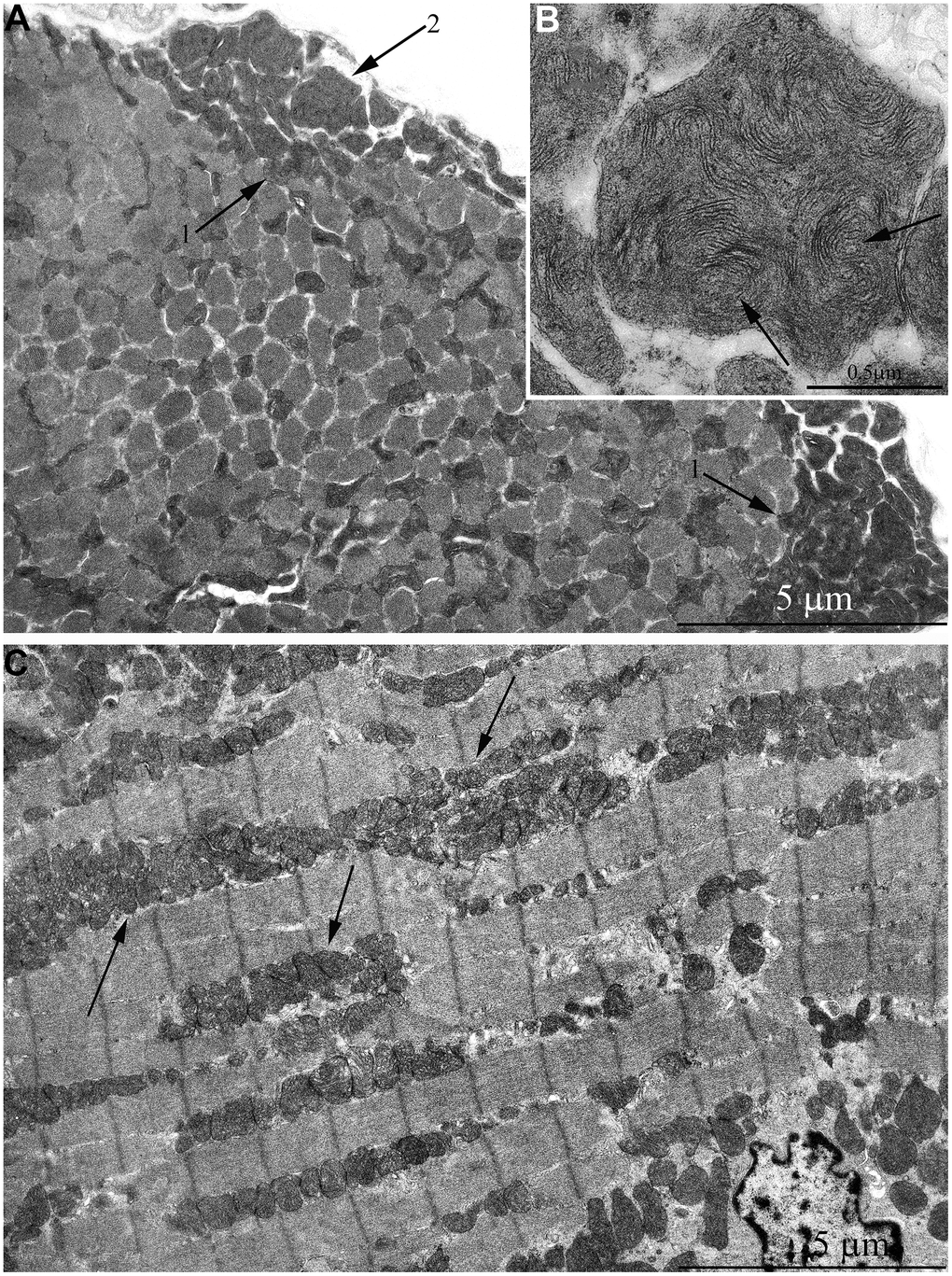
Figure 5.Ultrastructure of mitochondria in skeletal muscle of 7-year-old naked mole rat. (A) Cross section of muscle fiber. Clusters of large mitochondria in the subsarcolemmal area are indicated by arrows 1. Mitochondrion of specific ultrastructure, which is demonstrated in Figure 5B, is indicated by arrow 2. (B) Mitochondrion which specific ultrastructure, cristae in form of curled, wave-like structures are indicated by arrows. (C) Longitudinal section of muscle fiber, large clusters of mitochondria localized along myofibrils are indicated by arrows.