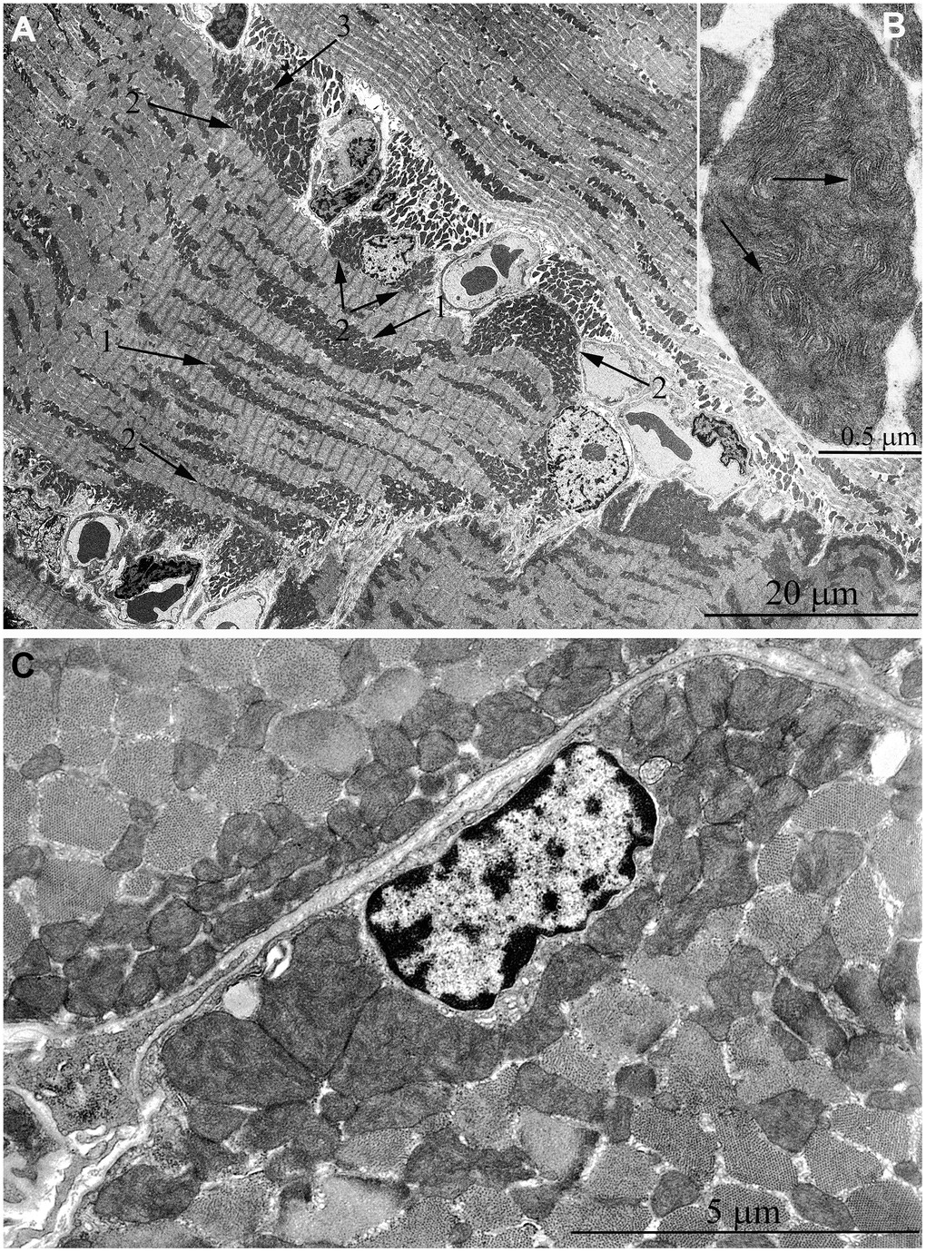
Figure 6.Ultrastructure of mitochondria in skeletal muscle of 11-year-old naked mole rat. (A) Longitudinal section of muscle fiber. Arrows 1 indicate large clusters of mitochondria located along myofibrils; arrows 2 indicate large clusters of mitochondria in the perinuclear and subsarcolemmal areas. Mitochondrion which specific ultrastructure is demonstrated under higher magnification in Figure 6B is indicated by arrow 3. (B) Mitochondrion which specific ultrastructure, convoluted stacks of cristae are indicated by arrows. (C) Cross section of muscle fiber. Similar ultrastructural pattern in the cross-section of muscle fiber is typical of cross-sections of cardiomyocytes, excluding subsarcolemmal localization of the nucleus.