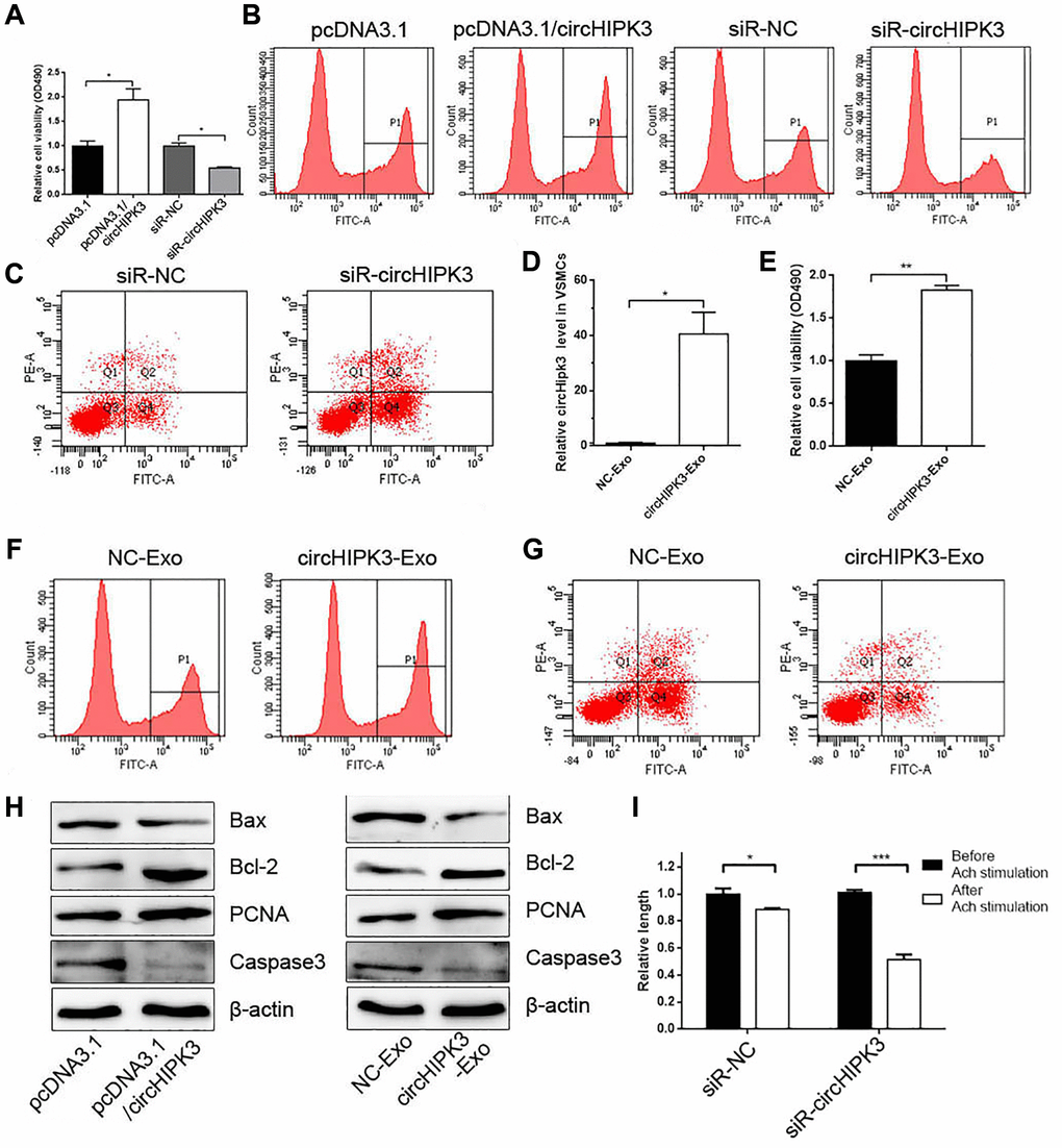
Figure 3.MAEC-derived exosomal circHIPK3 promotes VSMC proliferation and inhibited VSMC apoptosis. (A) CCK-8 was used to detect cell viability in VSMCs when overexpressing or knocking down circHIPK3 in VSMCs (*p < 0.05 pcDNA3.1/circHIPK3 vs. pcDNA3.1, *p < 0.05 siR-circHIPK3 vs. siR-NC). (B) Edu assay was used to detect cell viability by FCM. (C) FCM detected the apoptosis of VSMCs transfected with siR-circHIPK3 or siR-NC. (D) The relative expression of circHIPK3 in VSMCs incubated with exosomes isolated from 293A cells overexpressing circHIPK3 (*p < 0.05 NC-Exo vs. circHIPK3-Exo). (E) CCK-8 was used to detect cell viability in VSMCs incubated with NC-Exo or circHIPK3-Exo (**p < 0.01 NC-Exo vs. circHIPK3-Exo). (F) Edu assay was used to detect cell viability by FCM. (G) FCM detected the apoptosis of VSMCs. (H) Western blot analysis detected the expression of Bax, Bcl2, PCNA, and Caspase 3. (I) Cell length was calculated after infection with siR-NC or siR-circHIPK3 before and after inducing by Ach (*p < 0.05 Before Ach stimulation vs. After Ach stimulation for siR-NC, ***p < 0.001 Before Ach stimulation vs. After Ach stimulation for siR-circHIPK3).