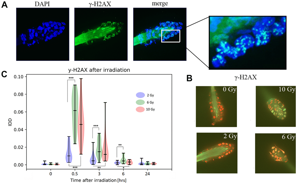
Figure 5.Immunofluorescent detection of γ-H2AX in murine hair follicles. (A) Representative images of a follicle after ionizing irradiation, extracted as maximum intensity projection from z-stack scanning by confocal spinning disc microscopy. Nuclei stained with DAPI and γ-H2AX foci and hair shaft autofluorescence are visible in the green channel. (B) Representative images used in the computational analyses of one z-stack layer, the red color of the nuclei (DNA) was selected artificially to maximize the visibility of green γ-H2AX foci. (C) Results of image analyses in terms of IDD (intensity of DNA damage), with each data point representing the area of the γ-H2AX signal related to the area of the nuclei in one scanned hair follicle in z-stack mode. Probability density of the data, maximal and minimal values, medians, and results of a multiple Kruskal-Wallis test (*p<0.05, **p<0.01, ***p<0.001) are showed in violin plots.