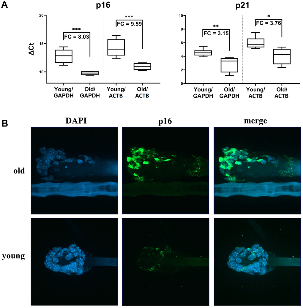
Figure 6.Markers of senescence analysis in hair follicular cells. (A) p16 and p21 gene expression (mRNA level) in hair follicles from young mice (6 months old) and old mice (2.5-3 years old) in boxplot graphs of ΔCt values normalized using GAPDH or ACTB. Graphs show medians, first quartile data distribution, minimal and maximal points, and fold change values as a specifying detail. Results of a t-test are also shown (*p<0.05, **p<0.01, ***p<0.001). (B) Representative images showing the higher p16 protein levels in murine hair follicles of old animals relative to those of young animals. Images were obtained from z-stack scanning with a confocal spinning disc microscope. DAPI-stained nuclei are shown in the blue channel and p16 signals in the green channel. Autofluorescence can be seen in a hair shaft. Magnification 60x objective with oil immersion.