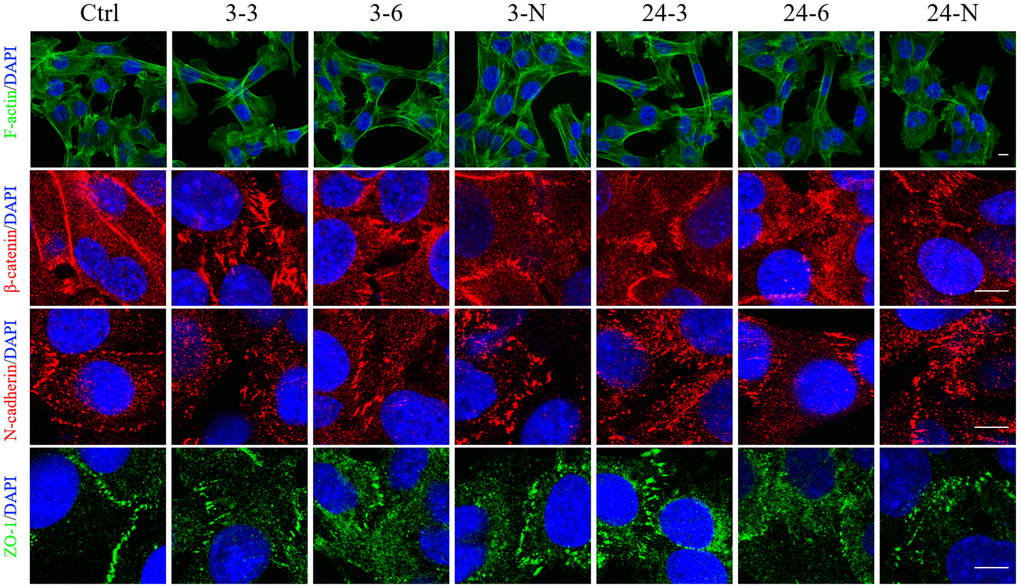
Figure 4.The spatial distributions of F-actin and BTB-associated proteins in TM-4 cells treated with two different concentrations of TiO2-NPs for 24 h. The distribution of intracellular microfilaments (F-actin) and the distribution of BTB-associated proteins: ZO-1 (green), N-cadherin (red), and β-catenin (red). “3–3”: 30 μg/ml, “3–6”: 60 μg/ml, “3-N”: TM-4 cells were treated with 5 mM NAC for 2 h and then treated with 60 μg/ml 3-nm TiO2-NPs for 24 h. “24–3”: 30 μg/ml, “24–6”: 60 μg/ml, "24-N”: TM-4 cells were treated with 5 mM NAC for 2 h and then treated with 60 μg/ml of 24-nm TiO2-NPs for 24 h. Scale bar=10 μm.