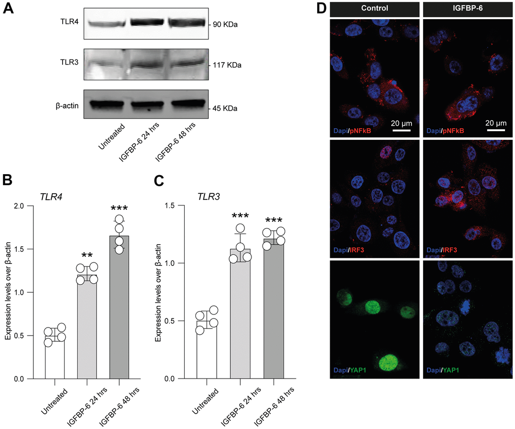
Figure 5.IGFBP-6 induces TLR4 signalling on HS5 cells. (A) HS5 cells exposed to 200 ng/mL of IGFBP-6 for 24h and 48h were lysed and subjected to immunoblotting using specific antibodies against TLR4 and TLR3. Protein content was normalized to the housekeeping protein β-actin. The entire assays were made in triplicate, a representative one is shown. Signals from immunodetected bands were semi-quantified by densitometry. (B, C) Statistical analysis of data revealed that TLR4 (B) and TLR3 (C) expression were significantly increased in the HS5 cells IGFBP-6- induced for 24h and 48h. Data are presented as means ± sem. **p < 0.01 and ***p < 0.001 vs. untreated. (D) Immunofluorescence analysis were performed on HS5 cells treated with IGFBP-6 at the final concentration of 200 ng/mL, followed by fixing and staining with anti-pNF-kB (red), anti-IRF3 (red), and anti-YAP1 (green). Nuclei were visualized using DAPI. Immunoreactivity was evaluated considering the signal-to-noise ratio of immunofluorescence (scale bar 20 μm).