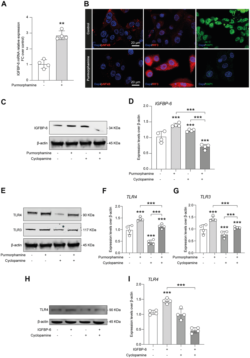
Figure 6.IGFBP-6-induced TLR4 signalling is controlled by SHH signalling through SMO. (A) qPCR results obtained for IGFBP-6 in HS5 cells exposed or not to purmorphamine. Relative mRNA expression level normalized with β-actin by using a comparative 2-ΔΔCt method. **P < 0.01 and ***P < 0.001. (B) Immunofluorescence analysis were performed on HS5 cells exposed or not to purmorphamine, followed by fixing and staining with anti-pNF-kB (red), anti-IRF3 (red), and anti-YAP1 (green). Nuclei were visualized using DAPI. Immunoreactivity was evaluated considering the signal-to-noise ratio of immunofluorescence (scale bar 20 μm). (C) HS5 cells exposed to purmorphamine, cyclopamine, or both were lysed and subjected to immunoblotting using a specific antibody against IGFBP-6. Protein content was normalized to the housekeeping protein β-actin. The entire assay was made in triplicate, a representative one is shown. Signals from immunodetected bands were semi-quantified by densitometry. (D) Statistical analysis of data revealed that IGFBP-6 expression was significantly increased after exposure to purmorphamine. Data are presented as means ± sem. **p < 0.01 and ***p < 0.001 vs. untreated. (E) HS5 cells exposed to purmorphamine, cyclopamine, or both were lysed and subjected to immunoblotting using specific antibodies against TLR4 and TLR3. Protein content was normalized to the housekeeping protein β-actin. The entire assay was made in triplicate, a representative one is shown. Signals from immunodetected bands were semi-quantified by densitometry. (F, G) Statistical analysis of data revealed that purmorphamine was able to increase while cyclopamine was able to suppress both TLR4 (F) and TLR3 (G) protein expression levels. Co-treatment with both SMO agonist and antagonist did not affect TLR4 and TLR3 expression levels, as compared to control cell cultures. Data are presented as means ± sem. **p < 0.01 and ***p < 0.001 vs. untreated. (H) HS5 cells exposed to IGFBP-6, cyclopamine, or both were lysed and subjected to immunoblotting using a specific antibody against TLR4. Protein content was normalized to the housekeeping protein β-actin. The entire assay was made in triplicate, a representative one is shown. Signals from immunodetected bands were semi-quantified by densitometry. (I) Statistical analysis of data revealed that TLR4 expression levels were significantly increased after IGFBP-6 stimulation, while a cotreatment with cyclopamine had a reducing effect on TLR4 expression. Data are presented as means ± sem. **p < 0.01 and ***p < 0.001 vs. untreated.