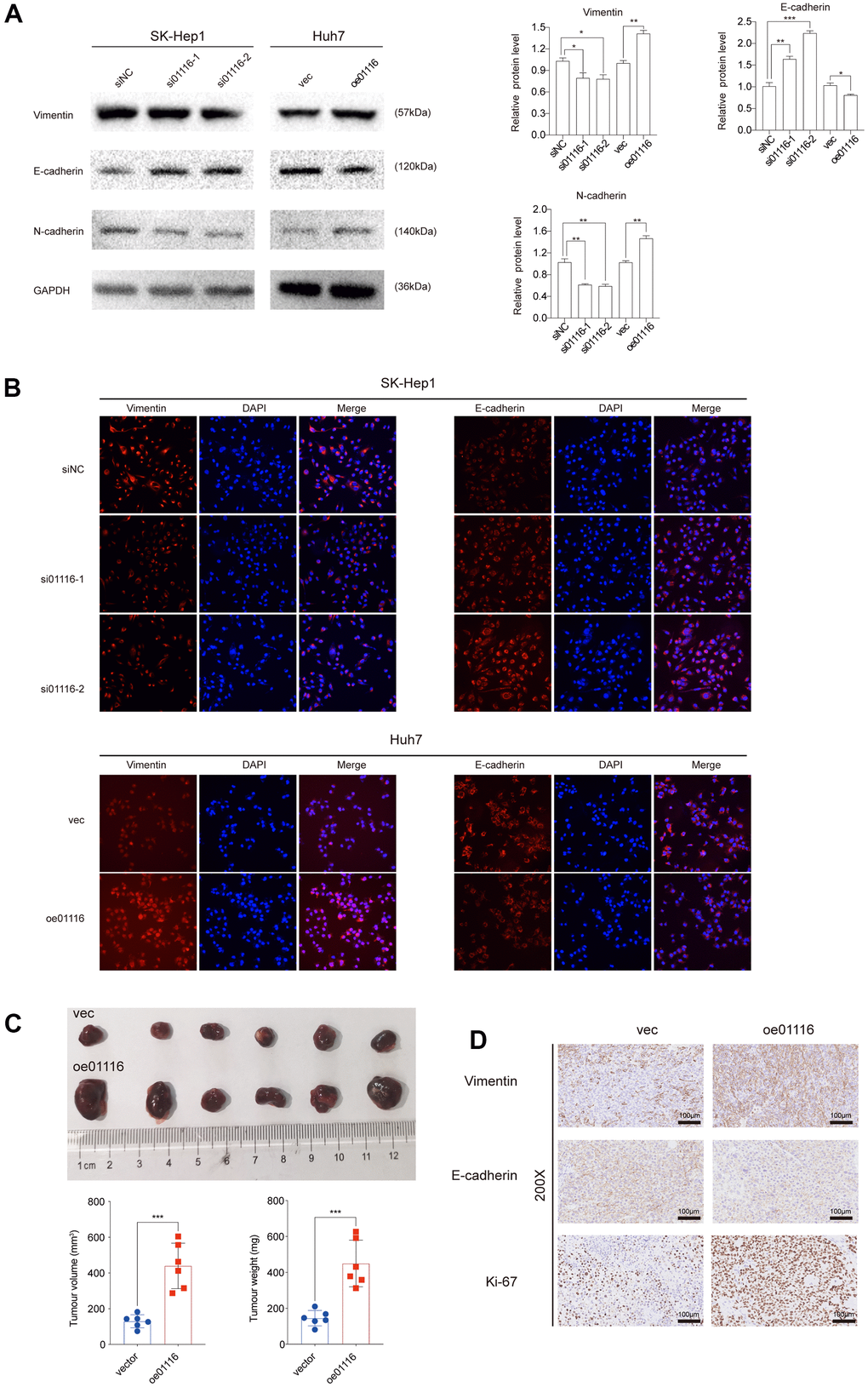
Figure 9.(A) EMT markers were examined via western blot analysis. (B) Immunofluorescence assay was used to detect EMT markers. (C) Image of subcutaneous tumor tissues. The volume and weight of tumors were measured. (D) Ki67, vimentin and E-cadherin were observed in subcutaneous tumor tissues by IHC. **P < 0.01 and ***P < 0.001.