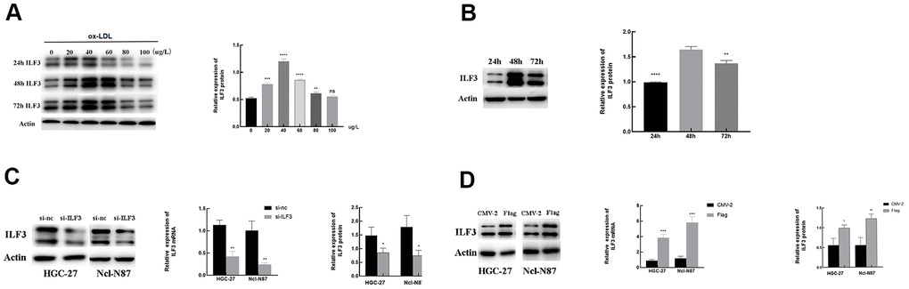
Figure 5.The relationship of the expression of ILF3 and ox-LDL. (A, B) ox-LDL promoted the expression of ILF3 in a time-concentration-dependent manner, the optimal concentration and intervention time was 40 μg/ml and 48h. (C) ILF3 was knocked down by ILF3-specific small interference RNA (siRNA) in HGC-27 and Ncl-N87 cells. The mRNA and protein expression level of ILF3 was verified by RT-qPCR and western blot. The ILF3 expression at the mRNA and protein levels was significantly lower in the ILF3-siRNA group compared with the negative control group in HGC-27 and Ncl-N87 cells. (D) ILF3 was overexpressed by ILF3-overexpressed plasmids (flag-ILF3) in HGC-27 and Ncl-N87 cells. The mRNA and protein expression level of ILF3 was verified by RT-qPCR and western blot. The ILF3 expression at the mRNA and protein levels was significantly higher in the flag-ILF3 group compared with the negative control group in HGC-27 and Ncl-N87 cells. **P < 0.01, ***P < 0.001, ****P < 0.0001 vs. 0 μg/L or 48 h groups.