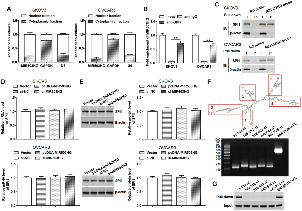
Figure 3.MIR503HG interacted with SPI1. (A) MIR503HG expression in cytoplasm and nucleus was evaluated in SKOV3 and OVCAR3 cells. U6 was used as a nuclear marker, and GAPDH was used as a cytoplasmic marker. (B) RIP assay was conducted with SPI1 antibody in SKOV3 and OVCAR3 cells, and anti-IgG was used as an internal control. (C) RNA pull-down experiment was performed by using biotinylated MIR503HG transcripts. (D, E) SPI1 mRNA and protein levels were determined after transfection with pcDNA-MIR503HG, MIR503HG siRNA or respective controls. (F) RNAfold web server was used to predict the secondary structure of MIR503HG, and MIR503HG sequence was divided into 5 truncated parts according to its secondary structure. (G) RNA pull-down assay was carried out with biotinylated MIR503HG truncated fragments. **P < 0.01. n = 6 in each group. Each test was repeated at least three times independently.