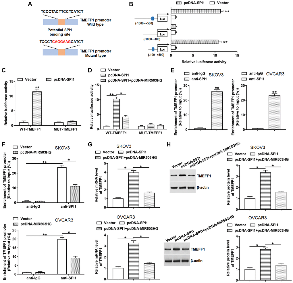
Figure 4.MIR503HG restrained SPI1-mediated transcriptional activation of TMEFF1. (A) ChIPBase database was used to predict the binding motif of SPI1 in TMEFF1 promoter region, and the mutation sites were shown in red. (B) Double luciferase reporter gene assay was performed using different truncated sequences of TMEFF1 promoter region. (C) WT-TMEFF1/MUT-TMEFF1 reporters were transfected into HEK293T cells together with pcDNA-SPI1/control vector, and the relative luciferase activities were detected. (D) HEK293T cells were transfected with WT-TMEFF1/MUT-TMEFF1 reporters together with pcDNA-SPI1 or pcDNA-SPI1 + pcDNA-MIR503HG, and the relative luciferase activities were analyzed. (E) ChIP assay was carried out to verify the binding of SPI1 to the TMEFF1 promoter region in SKOV3 and OVCAR3 cells. (F) ChIP assay was conducted with anti-SPI1 or anti-IgG in SKOV3 and OVCAR3 cells transfected with control vector or pcDNA-MIR503HG. (G, H) pcDNA-SPI1 was transfected into cells alone or together with pcDNA-MIR503HG, after transfection for 48 h, the mRNA and protein levels of TMEFF1 were measured. *P < 0.05, **P < 0.01. n = 6 in each group. Each test was repeated at least three times independently.