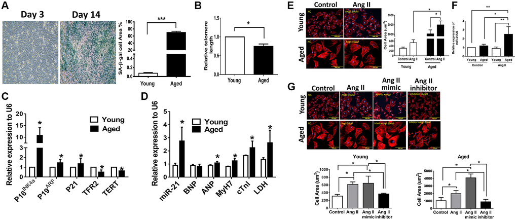
Figure 2.Angiotensin II (Ang II)-induced more cardiac hypertrophy and miR-21 expression in primarily isolated aged cardiomyocytes than in primary young cardiomyocytes. (A) The cultured neonatal rat cardiomyocytes for 14 days displayed cardiomyocytes senescence. SA-β-gal staining results for cardiomyocytes of neonatal rats. Blue precipitation in the cytoplasm was observed in the senescent cells. Percentage of β-gal-positive cardiomyocytes was increased in cultured cardiomyocytes for 14 days. (B) Telomere length expression in cardiomyocytes of neonatal rats. (C) The expression of cell senescence-associated protein in cultured neonatal rat cardiomyocytes was detected by qRT-PCR. (D) The levels of miR-21 and cardiac injury-associated genes in cultured neonatal rat cardiomyocytes were detected by qRT-PCR. (E) Immunofluorescence assay of F-actin was performed to identify the cell area in each group. Bar charts showing the individual cardiomyocyte cell areas. (F) The levels of miR-21 in cultured young and aged rat cardiomyocytes with and without treatment of Ang II detected by qRT-PCR. (G) Primarily isolated young and aged cardiomyocytes were transfected with a miR-21 mimic or inhibitor for 24 hours. Representative merged images of F-actin immunofluorescence staining of cardiomyocytes. Overexpression of miR-21 enhanced Ang II-induced cardiac hypertrophy, especially in primarily isolated aged cardiomyocytes. *P < 0.05, **P < 0.01, and ***P < 0.001 for difference from each group (N = 6–8).