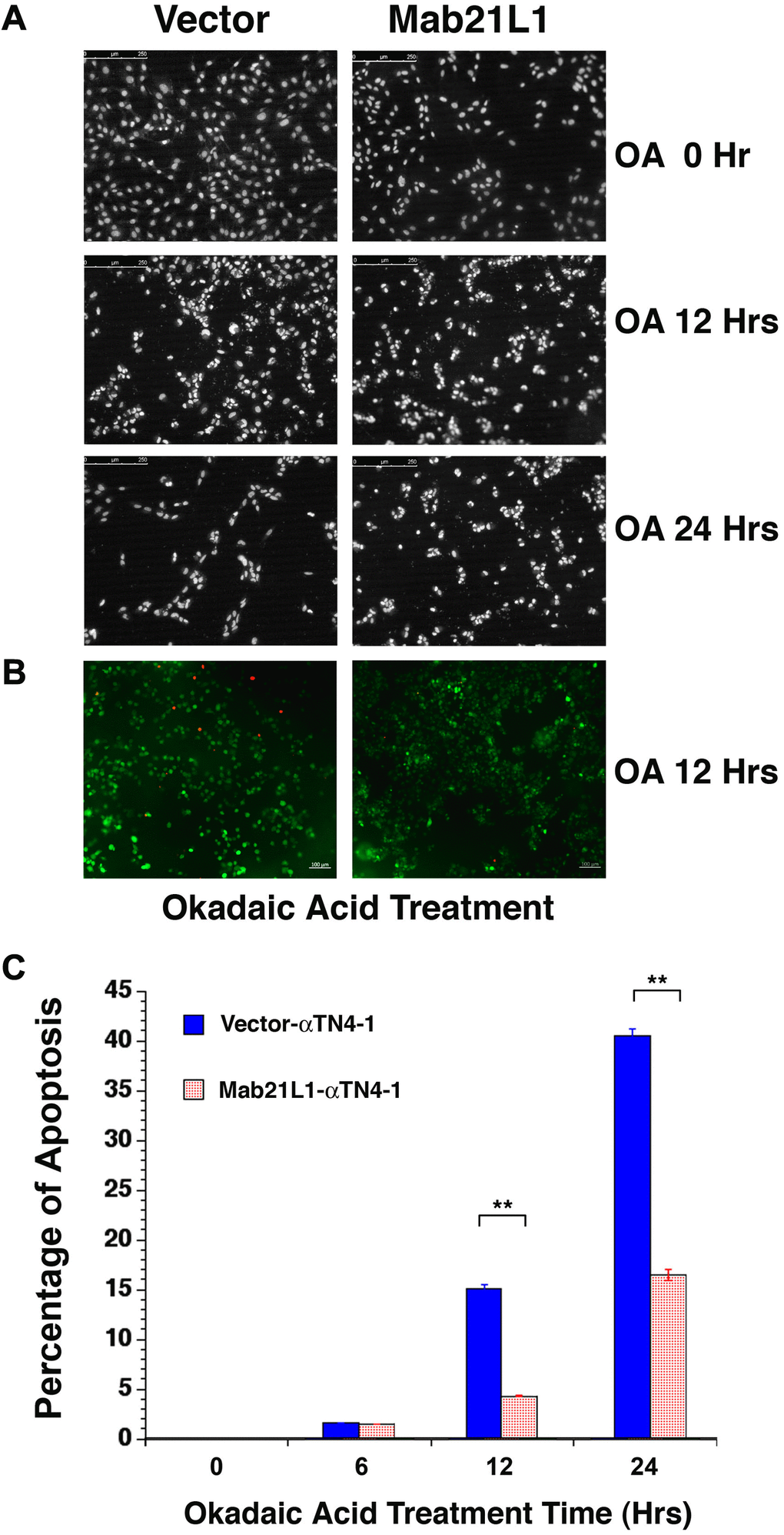
Figure 2.Analysis of okadaic acid (OA)-induced apoptosis of vector-αTN4-1 (vector) and MAB21L1-αTN4-1 (Mab21L1) cells. (A) Hoechst staining of OA-treated vector-αTN4-1 and MAB21L1-αTN4-1 cells for 0 to 24 hours. The apoptotic cells displayed fragmented or condensed nuclei, or were detached from the culture dishes, so that empty space appeared. Both vector-αTN4-1 and MAB21L1-αTN4-1 cells were grown to the density as shown in row one of Figure 2A (approximately 90% confluence, 0 Hr treatment), then subjected to 100 nM okadaic acid (OA) treatment with for 12 and 24 hrs. After OA treatment, the cells were processed for Hoechst staining as described (Mao et al., 2001). (B) Image of live cells (green color) and apoptotic cells (read color) of the vector-αTN4-1 and MAB21L1-αTN4-1 cells after 12 Hrs treatment by OA. (C) Quantitative results of apoptosis rate using live/dead assay as described (Wang et al., 2021). OA treatment for 12 hrs and 24 hrs induced about 15% and 41% apoptosis in vector-transfected cells, respectively. In contrast, only about 5% and 16% apoptosis were detected in MAB21L1-transfected cells after 12h and 24h-treatment with 100 nM OA. Note that MAB21L1 displayed the anti-apoptotic ability in αTN4-1 cells. Scale bar, 250 μm. N=3. ** p<0.01.