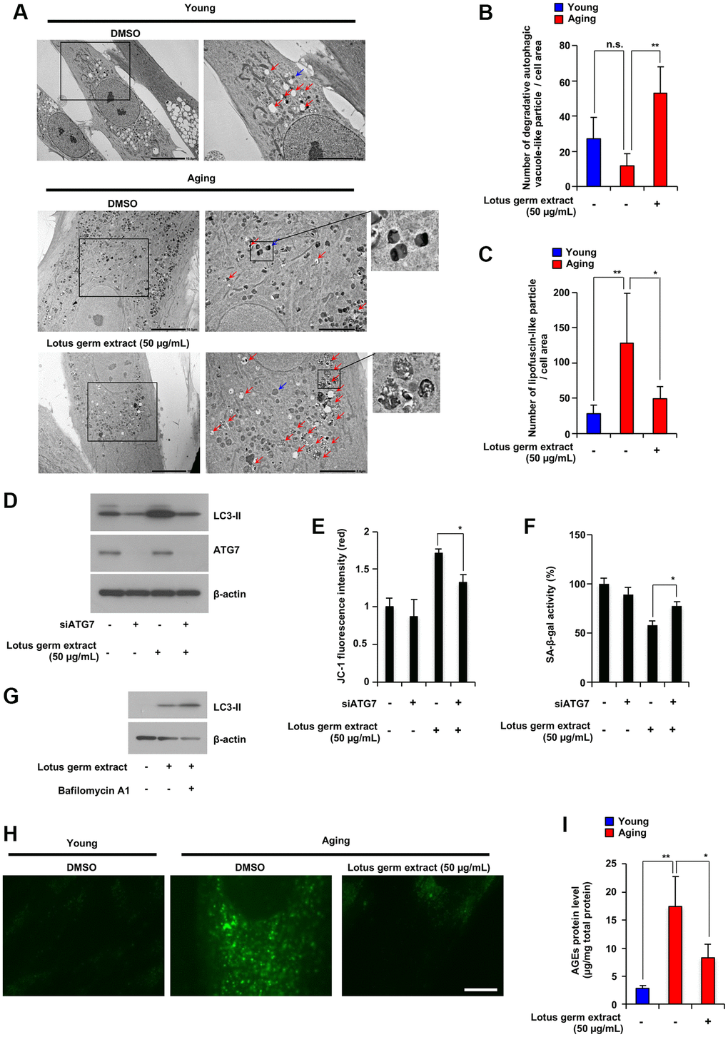
Figure 3.Lotus germ extract induced autophagy and decreased aging-related accumulation of lipofuscin-like particles and AGEs. (A–C) Young and aging NB1RGB cells with DMSO (−) or lotus germ extract (50 μg/mL) treatment (+) were subjected to TEM. The scale bar in the left panel represents 10 μm. Higher magnification images are shown in the right panel, with the scale bar representing 5 μm. Blue arrows indicate aged-related increased lipofuscin-like particles. Red arrows indicate degradative autophagic vacuole-like particles. The numbers of lipofuscin-like particles (B) and degradative autophagic vacuole-like particles (C) in each cell area were determined. Data are presented as the mean ± SD (n = 5). (D and E) Lotus germ extract induced autophagy, which plays an important role in the suppression of cellular aging phenotypes. Aging NB1RGB cells were transfected with siControl or siATG7 for 24 h, followed by treatment with DMSO (−) or 50 μg/mL lotus germ extract (+) for 24 h. Cells were subjected to immunoblotting using the indicated antibodies (D). JC-1 activity was determined based on fluorescence intensity (E). (F) Aging NB1RGB cells were transfected with siControl or siATG7 for 24 h, followed by treatment with DMSO (−) or 50 μg/mL lotus germ extract (+) for 3 days, and SA-β-gal activity was measured. (G) Treatment with the lotus germ extract stimulates autophagosome synthesis. Aging NB1RGB cells were treated with DMSO (−) or 50 μg/mL lotus germ extract (+) for 24 h, followed by treatment with or without bafilomycin A1 (1 μg/mL) (+) for 2 h. Cells were subjected to immunoblotting using the indicated antibodies. (H and I) Lipofuscin-like particle and AGE levels were decreased by lotus germ extract treatment. Young and aging NB1RGB cells were treated with DMSO (−) or 50 μg/mL lotus germ extract (+) for 3 days and subjected to fluorescence microscopy analysis to detect autofluorescence due to lipofuscin-like particles (H) or AGE levels by ELISA (I). Data are presented as the mean ± SD of three simultaneously performed experiments (E and F, I). P value was calculated using two-way ANOVA; n.s.: not significant, *P < 0.05, **P < 0.01.