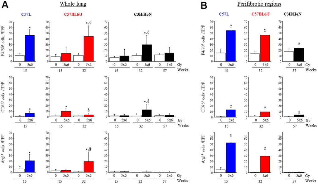
Figure 4.Accumulation of macrophages in irradiated lungs from three strains of mice. Immunohistochemical assays for (F4/80+), M1 macrophages (CD86+), and M2 macrophages (Agr-1+) were performed on lung tissue sections collected at the indicated timepoints after thoracic irradiation (5 x 6Gy). The numbers of cells were scored in whole lung (A) or within perifibrotic regions (within 400 μm of fibrotic regions) (B) in n=5 mice per time point and condition. Columns: mean, error bars: + SD, *p<0.05 for comparison to the corresponding 0 Gy by ANOVA with Tukey’s correction. §p<0.05 for comparison to lungs exposed to 5x6 Gy at 15 weeks by ANOVA with Tukey’s correction HPF: high power field (20X), IR: irradiation.