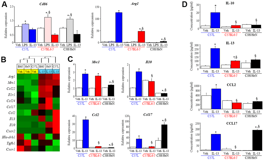
Figure 5.Characterization of macrophage phenotype across mouse strains after exposure to polarizing stimuli. Bone marrow derived macrophages from each strain were polarized with vehicle (PBS), LPS (1 ng/ml) or IL-13 (10 ng/ml). After 3 days of exposure to each stimulus, total RNA was isolated for further assays. (A) Polarization in response to LPS (M1) or IL-13 (M2) was evaluated by assessing the level of Cd86 and Arg1 mRNA with quantitative PCR (QPCR) normalized to β-actin mRNA. (B) The expression of genes related to M2 polarization was evaluated in macrophages treated with vehicle or IL-13 using the NanoString nCounter Gene Expression Assay and a custom code set. Unsupervised hierarchical clustering of M2 related genes was performed (left panel). (C) mRNA expression of Mrc1, Il10, Ccl2 and Ccl17 in polarized macrophages was confirmed by QPCR. (D) The concentrations of IL-10, IL-13, CCL2 and CCL17 in supernatants collected from polarized macrophages were determined with ELISA. Veh: PBS, BL6J: C57BL6/J, HeN: C3H/HeN. Columns: mean, error bars: +SD, *p<0.05 for comparison to the corresponding macrophages treated with vehicle. §p<0.05 for comparison to C57L macrophages exposed to IL-13 by ANOVA with Tukey’s correction.