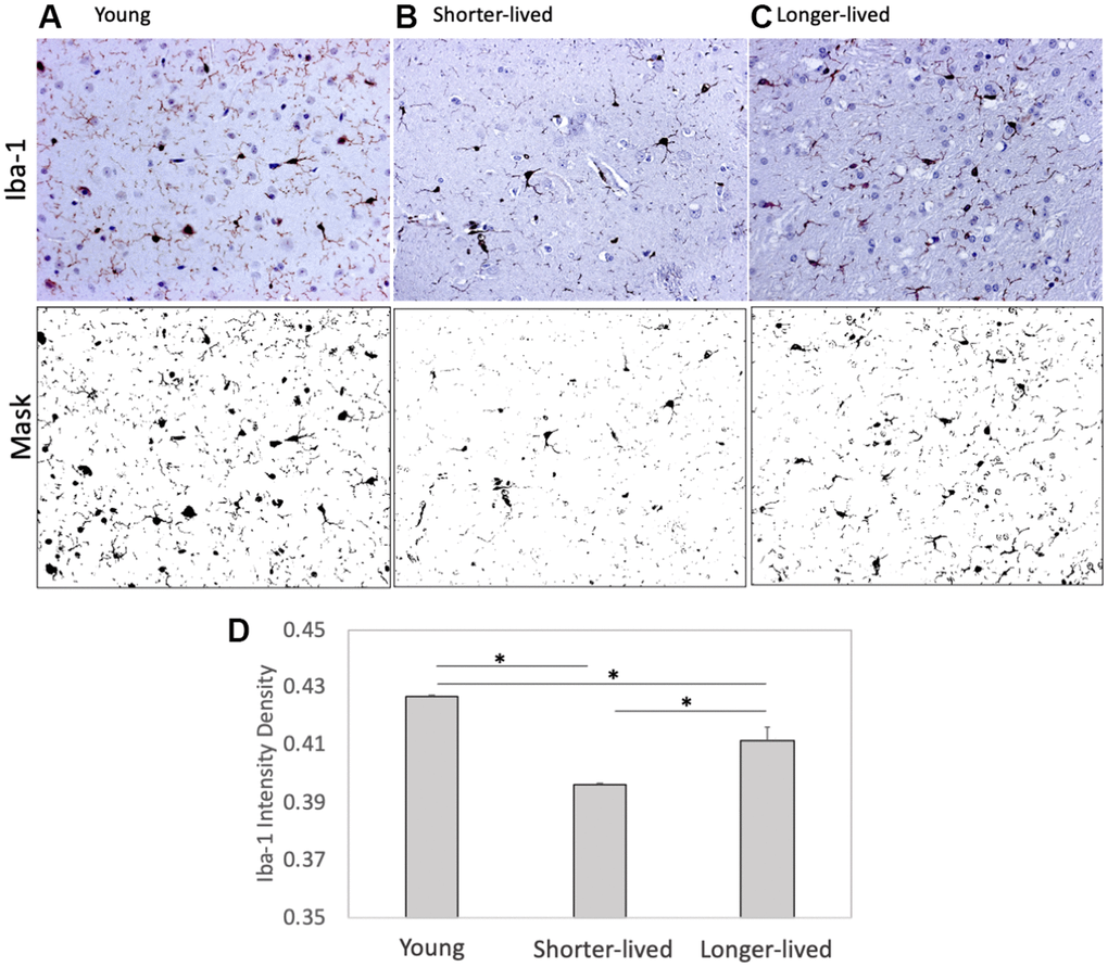
Figure 1.Expression of the microglia marker Iba1 in PFC from young animals and animals that lived to age. Paraffin-embedded tissue sections were stained with antibody anti- Iba-1 (AIF1, dark brown staining) and digitally imaged for density intensity quantification in whole section digital 8-bit images and binary masks using Image J (NIH). (A) Representative image taken from a young (4-7 yo) monkey, (B) representative shorter-lived (18-20 yo) group image, from an 18 yo monkey (M66), (C) representative longer-lived (23 – 29 yo) group image, from a 27 yo monkey (M208). (D) Iba-1 intensity density measured in ImageJ Fiji, using mask features. N=4/group. All representative images are 20X magnification. *p<0.0001 in multiple comparisons.