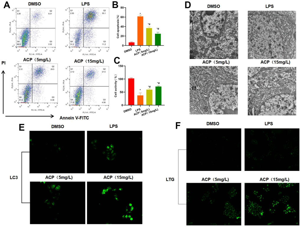
Figure 1.ACP suppressed cell injury and activated autophagy (n=3). (A, B) Flow cytometry revealed that LPS induced cell injury and significantly increased the apoptosis rate, while ACP suppressed cell apoptosis in a dose-dependent manner. *P<0.05 compared with DMSO, #P<0.05 compared with LPS. (C) Cell viability test suggested that cell viability decreased in LPS group, lower than that of DMSO group, while ACP suppressed LPS. *P<0.05 compared with DMSO, #P<0.05 compared with LPS. (D) Electron microscope observation demonstrated no obvious lysosome or autophagosome formation in DSMO, while weak autophagy activation was seen in LPS group, and obvious autophagosomes were detected in ACP, suggesting that ACP activated autophagy. (E) LC3 fluorescence staining revealed that LC3 was not significantly activated in DMSO, lowly expressed in LPS and significantly up-regulated in ACP, higher than that in DMSO and LPS group. (F) Lysosome probe LTG analysis suggested that ACP promoted lysosome formation and markedly enhanced the fluorescence intensity.