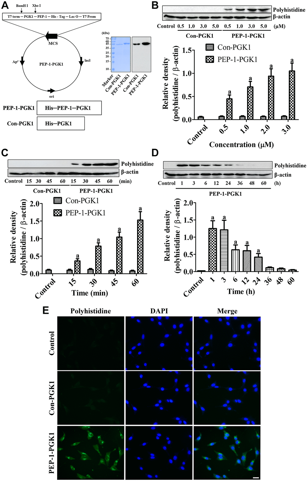
Figure 1.PEP-1-PGK1 and its control protein (Con-PGK1) are synthesized, and their expressions are confirmed, in HT22 cells by visualization of a polyhistidine tag. (A) PEP-1-PGK1 and Con-PGK1 are constructed, and their expressions are confirmed, in Escherichia coli cells. (B, C) Concentration- and time-dependent intracellular deliveries of Con-PGK1 and PEP-1-PGK1 are measured in HT22 cells after 3 μM of protein treatment and 1 h after the protein treatment, respectively. (D) Degradation of Con-PGK1 and PEP-1-PGK1 is assessed at various times after treatment. (E) Immunocytochemical staining visualizes the localization of delivered proteins in HT22 cells. Scale bar = 20 μm. (B–D) Optical densities of protein bands from western blotting are described as a value of polyhistidine/β-actin. Data are analyzed by a one-way analysis of variance, followed by a Bonferroni’s post-hoc test (ap < 0.05, significantly different from the control group). The bar graph represents the mean ± standard deviation.