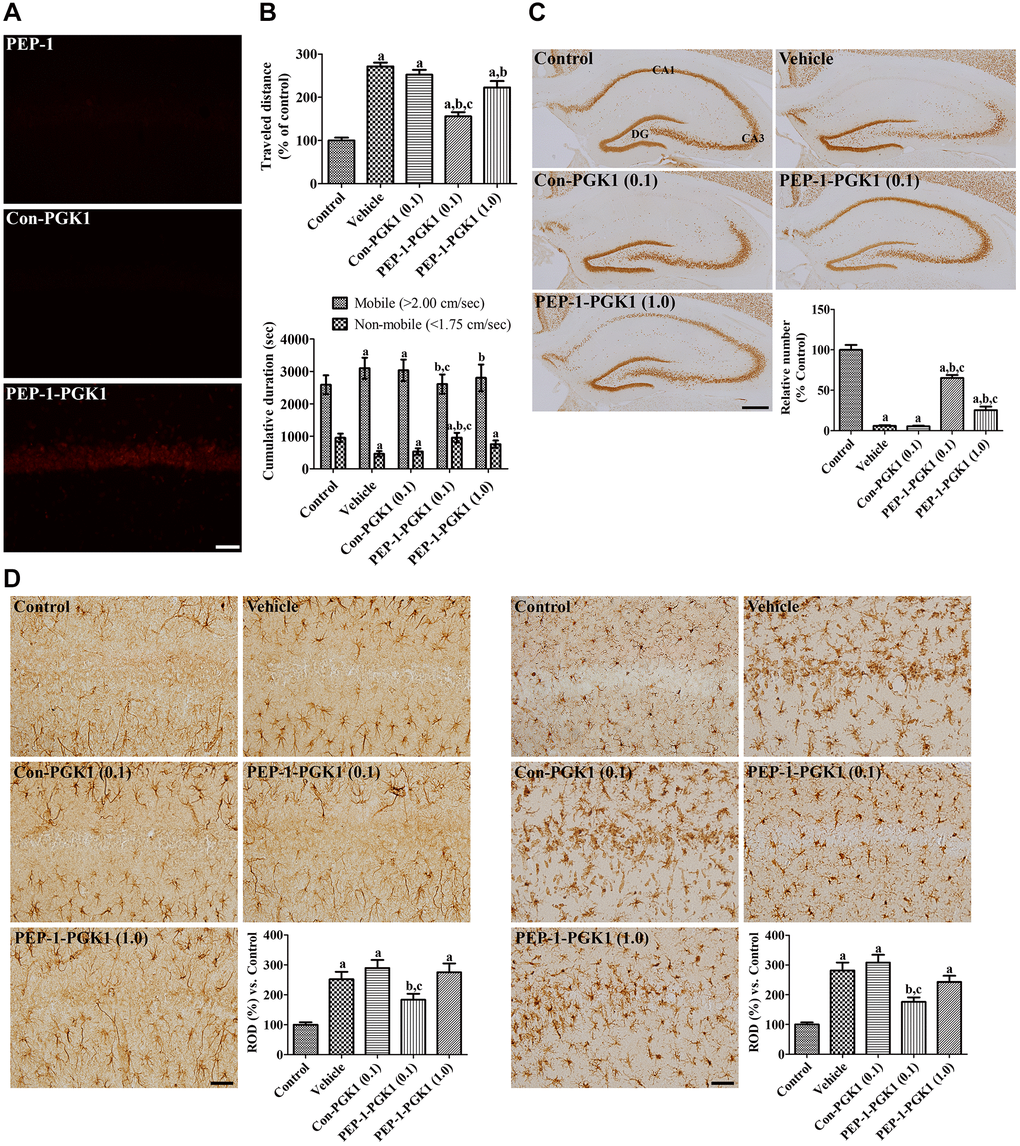
Figure 3.In vivo effects of Con-PGK1 and PEP-1-PGK1 on ischemia-induced damage in gerbils. (A) Immunohistochemical staining visualizes the delivery of proteins in the gerbil hippocampus. Scale bar = 50 μm. (B) Motor activities of gerbils are recorded, and the travel distance and time consumed 1 d after ischemia are reanalyzed. The travel distance is expressed as a percentile value versus the control group, and the time spent in mobile and non-mobile phases is also shown. (C) Surviving neurons are visualized by immunohistochemical staining for NeuN in the hippocampus 4 d after ischemia. NeuN-positive neurons are counted and demonstrated as a percentile value versus the control group. Scale bar = 400 μm. (D) GFAP-positive astrocytes and Iba-1-positive microglia are visualized in the hippocampal CA1 region 4 d after ischemia. Scale bar = 50 μm. Relative optical densities (ROD) are expressed as a percentage of the value of GFAP and Iba-1 immunoreactivity in the hippocampal CA1 region of the control group per section, respectively. (B–D) Data are analyzed by a one-way analysis of variance, followed by a Bonferroni’s post-hoc test (n = 6 per group; ap < 0.05, significantly different from the control group; bp < 0.05, significantly different from the vehicle-treated group; cp < 0.05, significantly different from the Con-PGK1-treated group). The bar graph represents the mean ± standard deviation.