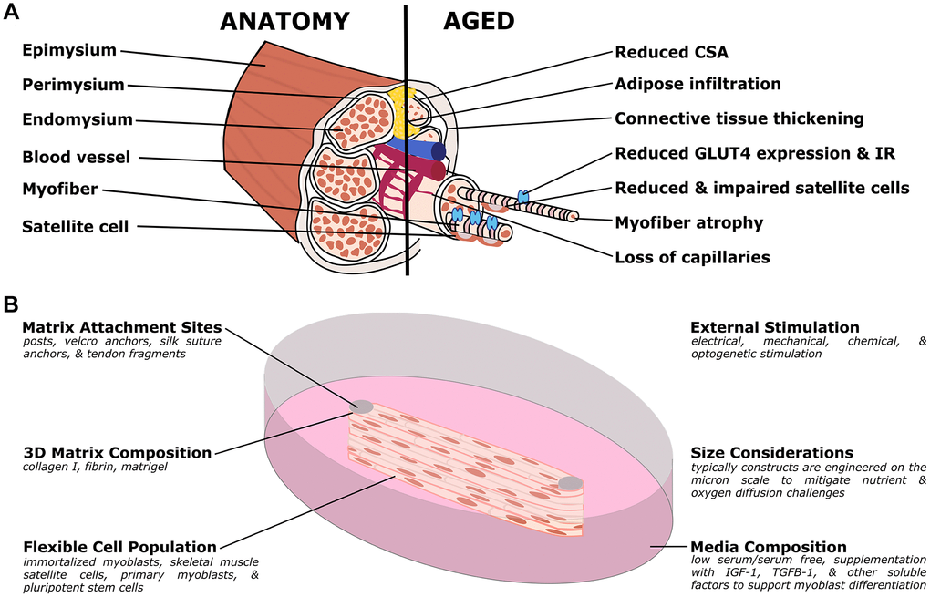
Figure 3.Organotypic models of skeletal muscle aging. (A) Simplified muscle anatomy and aging, focusing on the most commonly modeled components. The primary unit of muscle is the myofiber, a multinucleated cell responsible for contraction. Specialized matrix (endomysium, perimysium, and epimysium) support and organize the tissue. Satellite cells are an important stem cell population for the muscle, and the muscle is supported by a host of other cell types including nerves, fibroblasts, adipose, and vascular cells. In aged muscle, cross-sectional area (CSA) is reduced, in part due to myofiber atrophy, and decreasing capillary and satellite cell density. Conversely, there is increased infiltration of adipose and thickening of the connective tissues. At the molecular level, there is decreased expression of GLUT4, an important glucose transporter, and insulin resistance (IR) frequently develops. (B) Organotypic models of muscle have several unique challenges but have distinct advantages over other traditional models. Muscle cultures are contractile, and require anchoring to prevent collapse. Typical approaches include posts (although other methods are used) to provide points of resistance for the muscle to pull against. In order to study active contraction, researchers have used various stimulation methods, including electrical and optogenetic methods. Due to the high metabolic demand, the cultures are typically quite small, to allow nutrients and waste to diffuse more readily. As with other organotypic models, the matrix, cell population, and media can be customized for the research question.