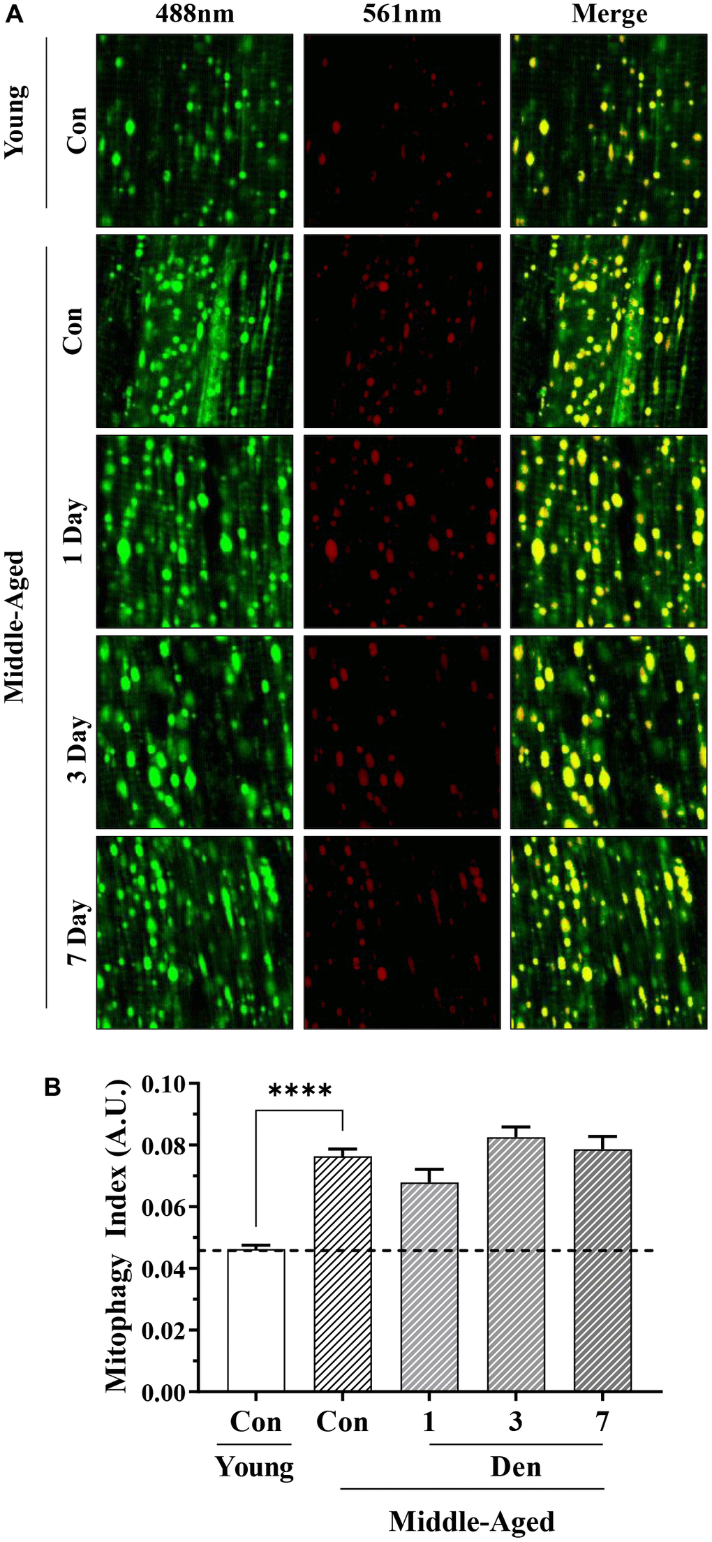
Figure 5.Assessment of mitophagy flux by mt-Keima fluorescence in young and middle-aged control and denervated skeletal muscle. (A) Representative fluorescence images from the TA muscle of mt-Keima in green (ex. 488nm; mitochondria), red (ex. 561nm; mitolysosomes), and merged. (B) Mitophagy index as calculated by red/total red+green fluorescence. ****p<0.0001, t-test between young and middle-aged control. 12 biological replicates were used for young and middle-aged control, 4 biological replicates were used for each denervation time-points. 8 images were taken per biological replicate (N=96/young and middle-aged control, N=32/denervation time point).