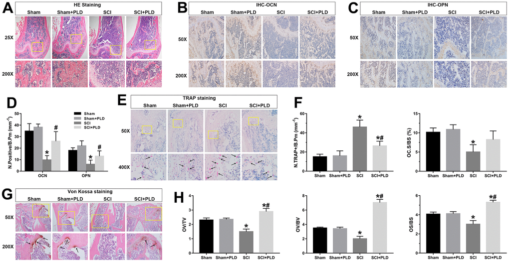
Figure 5.Effects of PLD on bone morphology, bone formation, calcium deposition and bone resorption. (A) Representative photomicrographs of H&E-stained sections. IHC analysis of (B) OCN and (C) OPN protein expression in distal femurs. (D) Number of OCN+ and OPN+ osteoblasts on the bone surface. (E) Representative images of TRAP-stained distal femoral sections. Arrows indicate TRAP+ cells. (F) Histo-morphometric quantification of osteoclast number per bone perimeter (N.TRAP+/B.Pm), and normal osteoclasts surface per bone surface (OC.S/BS). (G) Representative images of von Kossa-stained mouse femoral trabecular bone sections. Arrows indicate the presence of mineralized bone. (H) Quantitative analysis of osteoid volume versus total volume (OV/TV), osteoid volume versus bone volume (OV/BV), and osteoid surface versus bone surface (OS/BS). Measurements were presented as mean ± S.D.; n=6 to 7 per group; *P < 0.05, vs. the Sham group; #P < 0.05, vs. the SCI group.