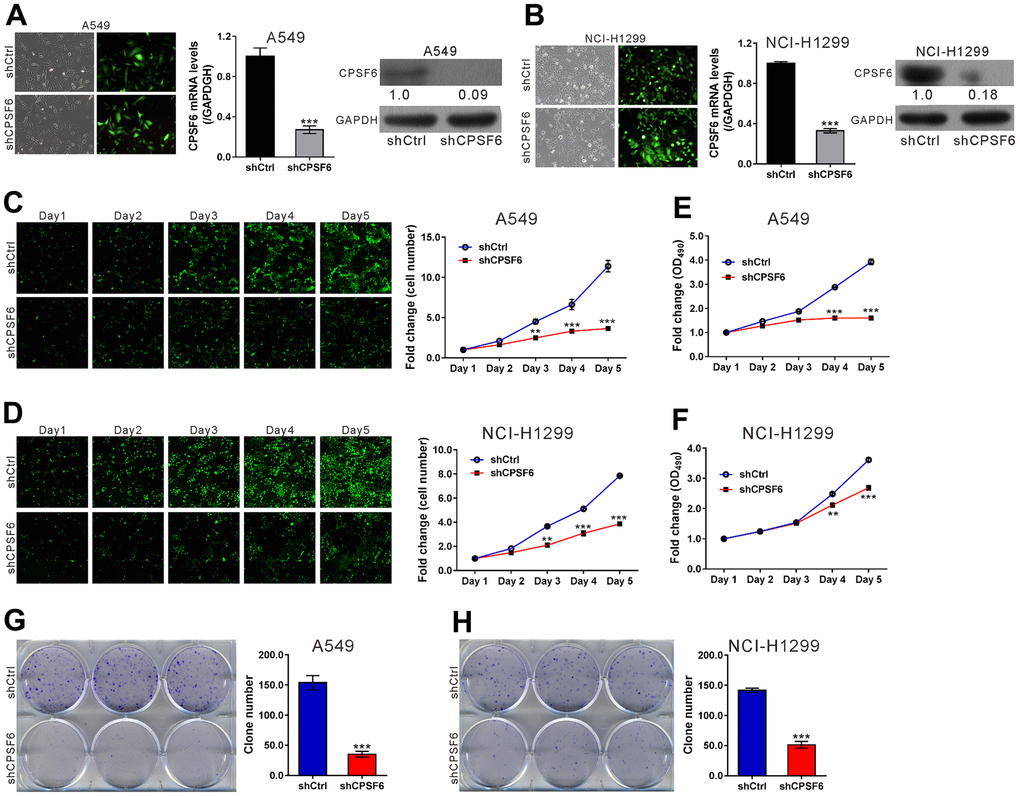
Figure 3.CPSF6 knockdown suppressed cell proliferation of LUAD cells. A549 and NCI-H1299 cells were transduced with lentivirus expressing CPSF6 shRNA (shCPSF6) or control shRNA (shCtrl). (A, B) At 72 h later, infection efficiency was estimated by counting the numbers of GFP expressing cells under a fluorescence microscopy. Magnification: 200× (left panel). Real-time PCR analysis and western blotting were conducted to assess mRNA (middle panel) and protein expression (right panel) of CPSF6, respectively. The densitometric analysis of western blotting was shown below the blot. (C, D) The Celigo Cell Counting assay was done for 5 days. The fold change of cell number was calculated relative to Day 1. (E, F) MTT assay was used to estimate cell proliferation. The fold change of OD490 was calculated relative to Day 1. (G, H) Colony formation assay was continued for 8 days to evaluate cell proliferation. **P<0.01, ***P<0.001 versus shCtrl. The number of cell experiments in parallel (n=3).