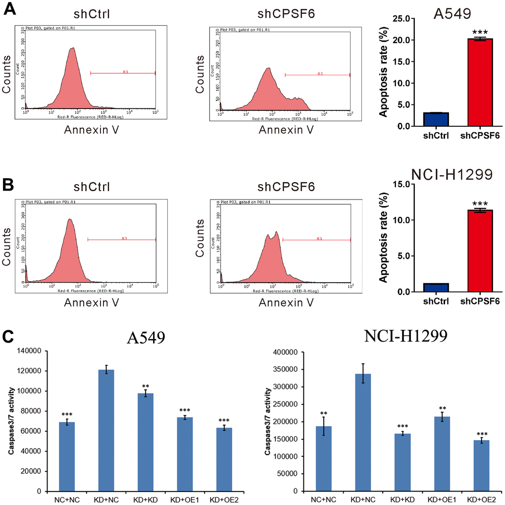
Figure 4.CPSF6 knockdown induced apoptosis of LUAD cells. A549 (A) and NCI-H1299 cells (B) were transduced with lentivirus expressing CPSF6 shRNA (shCPSF6) or control shRNA (shCtrl), labelled with the Annexin V Apoptosis Detection kit, and analyzed on a flow cytometry. Representative images and the apoptotic rates of three independent experiments are shown. ***P<0.001 versus shCtrl. (C). Caspase3/7 activity of A549 and NCI-H1299 cells were detected between five groups after transducing with lentivirus for 5 days. Control cell + control shRNA (NC+NC), CPSF6 shRNA + control shRNA (KD+NC), CPSF6 shRNA + GSK3B shRNA (KD+KD), CPSF6 shRNA + IRS1 Over Expressed (KD+OE1), CPSF6 shRNA + JUN Over Expressed (KD+OE2). **P<0.01, ***P<0.001 versus KD+NC. The number of cell experiments in parallel (n=3).