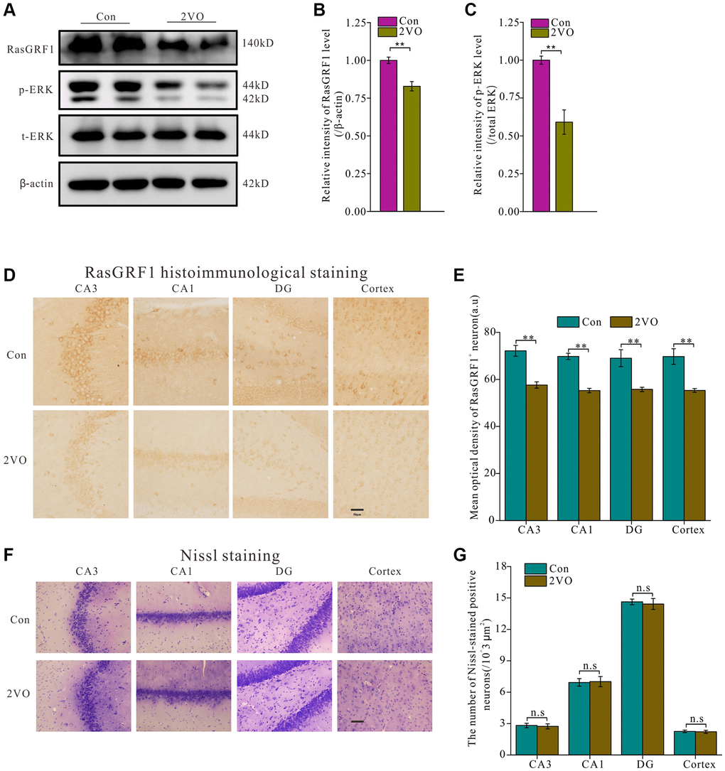
Figure 2.RasGRF1 expression in hippocampus dramatically decreased after chronic cerebral hypoperfusion. (A) The RasGRF1 expression, the total and phosphorylated ERK expression were assayed by Western blot. β-actin was as inner control for samples loading normalization; (Con (n = 3), 2VO (n = 3)). (B) The RasGRF1 relative expression level was calculated and analyzed statistically. (C) The p-ERK relative expression level was calculated and analyzed statistically. Relative intensity of p-ERK level is calculated by p-ERK/total ERK. Total ERK was as inner control for samples loading normalization. (D) RasGRF1 distribution in brain was observed with immunohistochemical staining; (Con (n = 1), 2VO (n = 1)). (E) In hippocampal subregions and cortex, the RasGRF1-positive cells were counted and analyzed. (F) The neurons number was observed with Nissl staining; (Con (n = 1), 2VO (n = 1)). (G) The Nissl-stained positive cells were counted and analyzed. Compared with Control, **p < 0.01. Scale bar = 50 μm. All of the experiments were repeated three times.