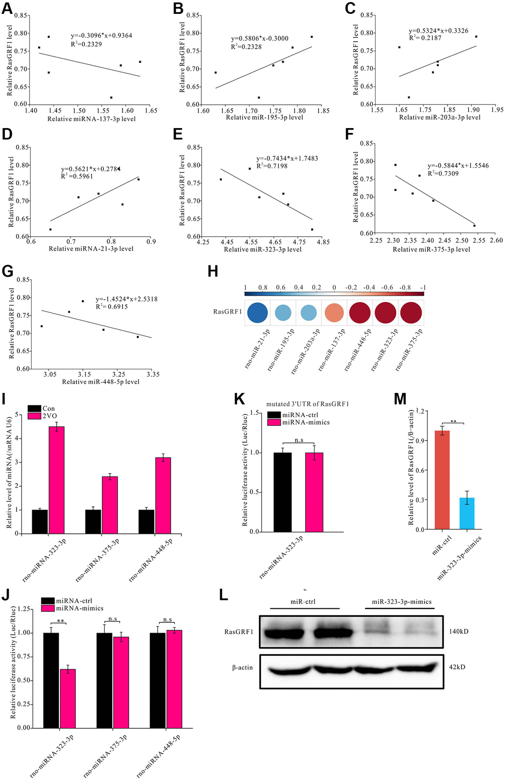
Figure 4.miR-323-3p could regulate the expression of RasGRF1 through binding to the 3′UTR. The total RNAs were extracted, and RasGRF1 and the predicted bind miRNAs levels were measured by RT-PCR. (A–G) The correlation between miRNAs (rno-miR-137-3p, rno-miR-195-3p, rno-miR-203a-3p, rno-miR-21-3p, rno-miR-323-3p, rno-miR-375-3p, rno-miR-448-5p) and RasGRF1 levels were analyzed. (H) The correlation degree was displayed with heatmap. (I) rno-miR-323-3p, rno-miR-375-3p and rno-miR-448-5p level in hippocampus after CCH were showed. (J) Dual luciferase reporter assayed the binding and expression regulation of targeted RasGRF1 mRNAs 3’UTR by miRNA mimics and negative control of miRNA-323-3p, miRNA-375-3p and miRNA-448-5p. (K) The binding and regulation between mutated 3’UTR of RasGRF1 mRNA and miRNA-323-3p was assayed by dual luciferase reporter. (L) RasGRF1 level after rno-miR-323-3p mimic treatment was detected by Western blot. β-actin was as inner control for samples loading normalization. (M) The RasGRF1 relative expression level was calculated and analyzed statistically. Compared with miRNA-Control, **p < 0.01. All of the experiments were repeated three times.