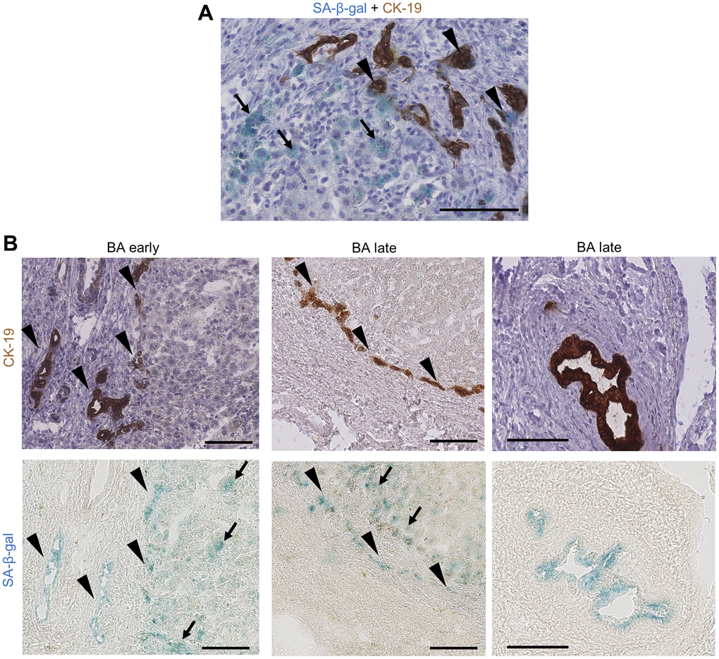
Figure 2.Cholangiocytes and perinodular hepatocytes display cellular senescence in BA livers. (A) Co-staining of SA-β-gal activity and CK-19 IHC on BA livers: some bile ductules cholangiocytes show staining co-localization (arrowheads) while perinodular hepatocytes are only positive for SA-β-gal (arrows); (B) Serial staining of SA-β-gal activity and CK-19 IHC on BA livers cryosections. Left and middle images: bile ductules (arrowheads) and perinodular hepatocytes (arrows) are positive for SA-β-gal in both early and late stage BA. Right images: remaining large septal bile duct display SA-β-gal activity. BA: biliary atresia; IHC: immunohistochemistry; SA-β-gal: senescence-associated beta-galactosidase. Scale bars = 100 μm.