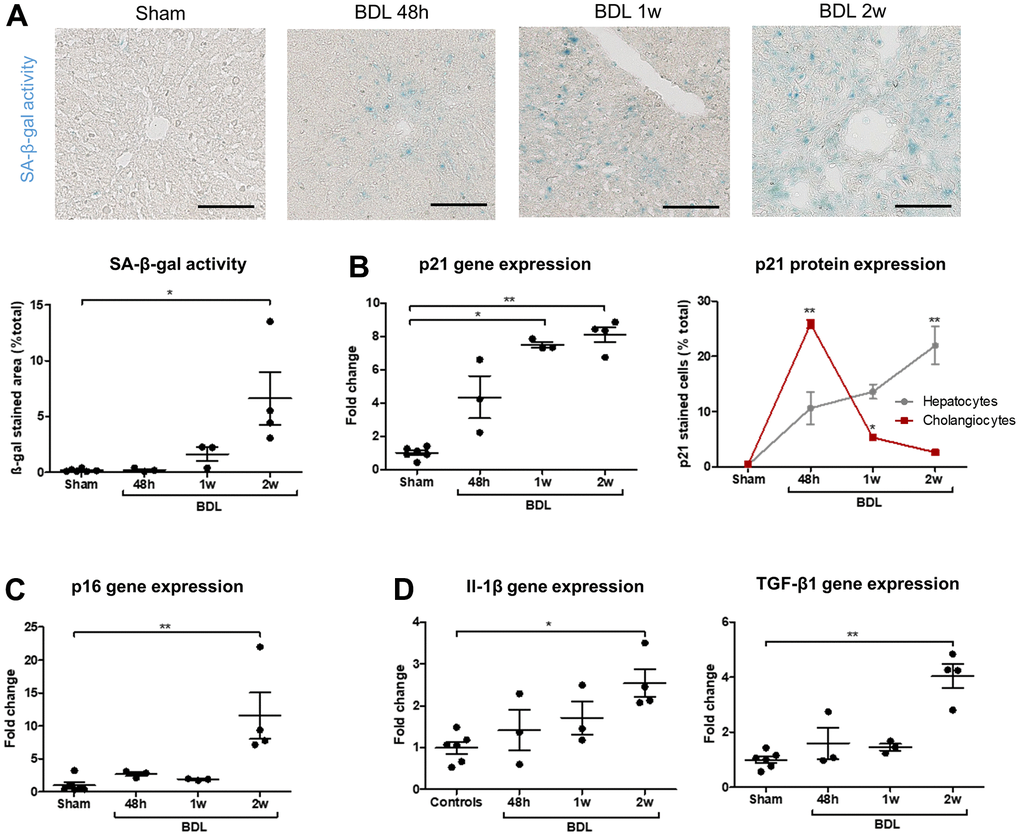
Figure 4.Senescence progressively develops in BDL rats and appears in cholangiocytes before hepatocytes. (A) SA-β-gal activity increases after the surgery. (B) p21 gene expression progressively increase in BDL livers as compared to controls. Senescence (p21-positive cell percentage) is maximal in cholangiocytes 48 hours post-BDL, while hepatocytes senescence increases progressively to become significant only two weeks after the surgery. The percentages of p21-positive cells at different timepoints are compared to the correspondent cellular type of control rats. (C) p16 gene expression increases in diseased rats two weeks post-surgery. (D) Gene expression of SASP markers Il-1β and TGF-β1 increase in BDL livers as compared to controls. BDL: bile duct ligation; SA-β-gal: senescence-associated β-galactosidase; SASP: senescence-associated secretory phenotype; 48h – 1w – 2w : rats sacrificed 48 hours (n=3) – 1 week (n=3) – 2 weeks (n=4) after BDL surgery. Data is presented as mean ± SEM; *p≤0.05; **p<0.01. Scale bars = 100μm.