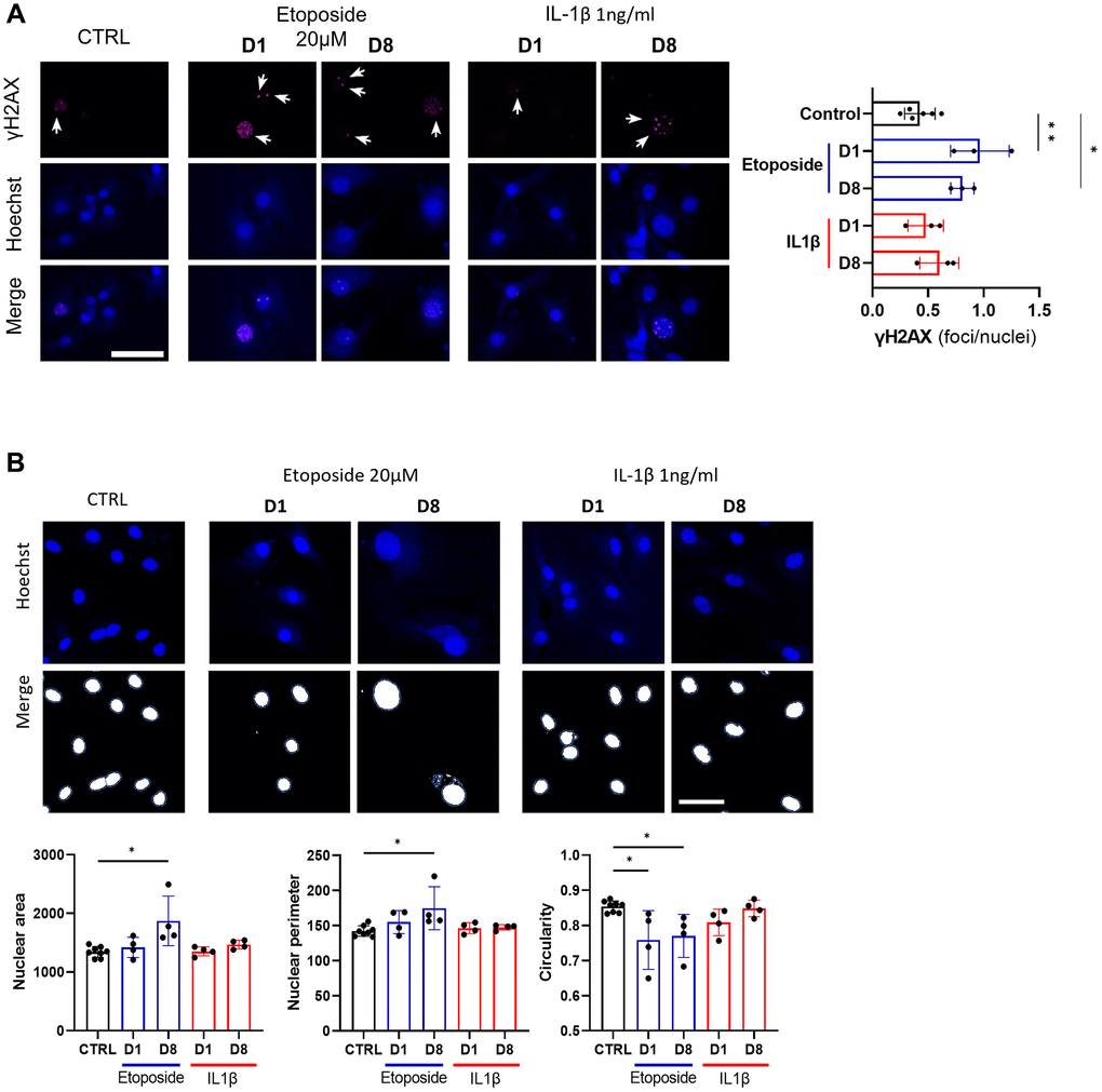
Figure 4.DNA damage assessment and senescence-associated nuclear features measure in Etoposide, and IL-1β treated HACs. HACs were treated with Etoposide 20 μM (blue) for 24 h or chronically with 1 ng/mL IL-1β (red) for the length of the experiment. (A) γH2AX immunofluorescence was used to identify DNA damage-associated foci and Hoechst staining to visualize the nucleus at day 1 and day 8. Quantification of the average number of foci per nuclei is shown. (B) Nucleus surface, perimeter and circularity was analyzed using CellProfiler software on the Hoechst channel. Scale bars = 50 μm. Data are shown as mean ± SD, (n = 3). P-values were calculated by Kruskal-Wallis test, *p ≤ 0.05; **p < 0.01.