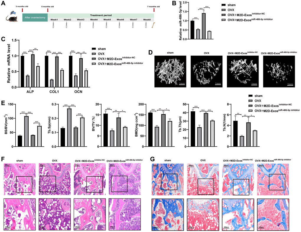
Figure 2.M2D-Exo-derived miR-486-5p accelerates bone formation in vivo. (A) Schematic diagram illustrating the experimental design. (B) qRT-PCR analysis of miR 486-5p levels in bone specimens from OVX mice after treatment with PBS, M2D-Exosinhibitor-NC, or M2D-ExosmiR-486-5p inhibitor. (C) qRT-PCR analysis of ALP, OCN, and COL1 mRNA levels in bone specimens from OVX mice after treatment with PBS, M2D-Exosinhibitor-NC, or M2D-ExosmiR-486-5p inhibitor. (D) Representative images showing the three-dimensional trabecular architecture in distal femurs determined by microCT reconstruction. Scale bars, 1 mm. (E) MicroCT measurements of BS/BV, Tb. Sp, BMD, BV/TV, Tb. N, and Tb. Th in the distal femurs of the OVX mice after treatment with PBS, M2D-Exosinhibitor-NC, or M2D-ExosmiR-486-5p inhibitor. (F) H&E staining indicates trabecular density. Scale bars indicated 200 μm and 500 μm. (G) Masson trichrome staining indicates trabecular density and collagen. Scale bars indicated 200 μm and 500 μm. n = 5 mice/group. Data are expressed as the mean ± SEM, *p < 0.05, **p < 0.01, ***p < 0.005.