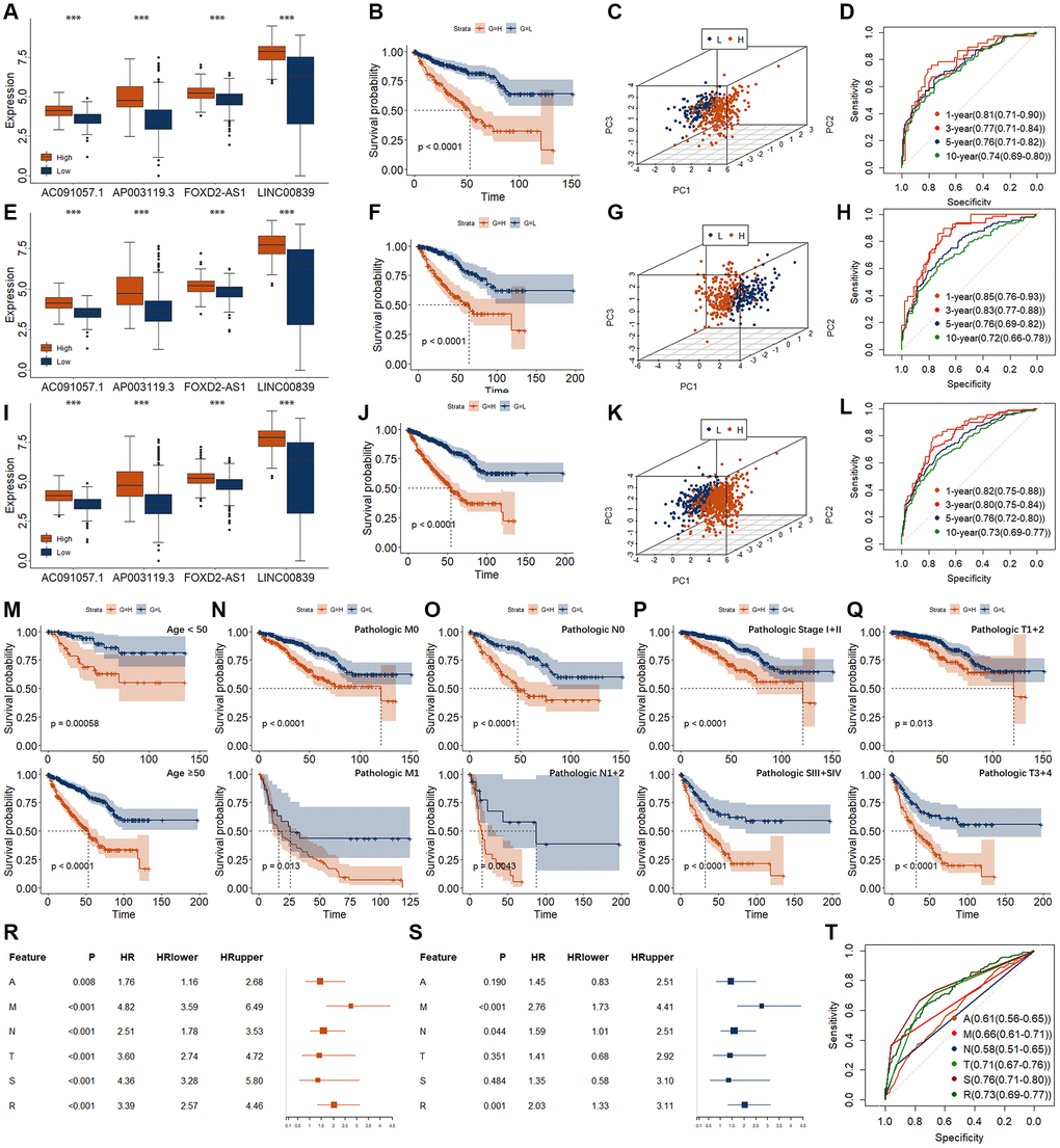
Figure 4.Establishment and validation of risk models for RCC. (A–D) Evaluation of risk model in training group, including expression level (A), K-M curve (B), PCA (C), and time-dependent ROC curve (D). (E–H) Evaluation of risk model in validation group, including expression level (E), K-M curve (F), PCA (G), and time-dependent ROC curve (H). (I–L) Evaluation of risk model in entire group, including expression level (I), K-M curve (J), PCA (K), and time-dependent ROC curve (L). (M–Q) K-M curves of risk models in entire group for different clinical phenotypes. (M) for age. Upper represents <50. Lower represents ≥50. (N) for pathologic M. Upper represents M0. Lower represents M1. (O) for pathologic n. Upper represents N0. Lower represents N1 + 2. (P) for pathologic T. Upper represents T1 + 2. Lower represents T3 + 4. (Q) for pathologic Stage. Upper represents SI + II. Lower represents SIII + IV. (R, S) Results of univariate (R) and multivariate (S) Cox regression for the risk model and different clinical feature. A, represents age. M, represents pathologic M. N, represents pathologic N. T, represents pathologic T. S, represents pathologic Stage. R, represents risk model. (T) ROC curve for the risk model and different clinical feature. *p < 0.05. **p < 0.01. ***p < 0.001.