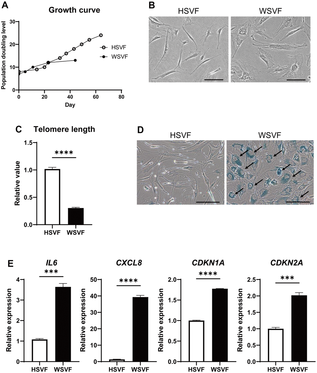
Figure 2.WSVF exhibits cellular senescence and increased expression levels of inflammatory genes. (A) Growth curves of SVF derived from a healthy individual and a patient with WS. (B) Comparison of the morphological features of the SVFs. Scale bar, 300 μm. (C) Quantification of the telomere length analyzed by quantitative real-time polymerase chain reaction. Data are presented as means ± S.E.M. of three technical replicates. For statistical analysis, student t-test was performed (****p < 0.0001). (D) Representative images of SA-β-gal staining of SVF. Black arrows indicate SA-β-gal-positive cells. Scale bar, 300 μm. (E) Quantitative real-time polymerase chain reaction of the relative expression of senescence-related genes. Data are presented as means ± S.E.M. of three technical replicates. For statistical analysis, student t-test was performed (ns, not significant; ***p < 0.001; ****p < 0.0001). WS: Werner syndrome; SVF: stromal vascular fraction; SA-β-gal: senescence-associated β-galactosidase.