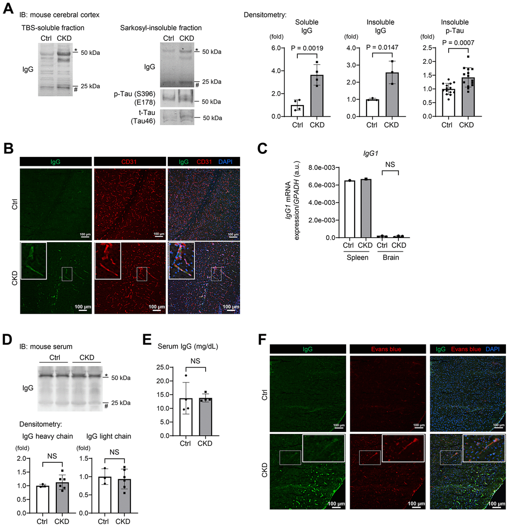
Figure 3.CKD increases a leakage of IgG from cerebral blood vessels to the brain parenchyma in mouse hippocampus and cerebral cortex. (A) Western blotting showed that CKD increased the expressions of soluble IgG (n = 4 per group), insoluble IgG (n = 3 per group), and phosphorylated tau (n = 14 per group) in the sarkosyl-insoluble (aggregated) fraction of the cerebral cortex. (B) The immunofluorescence study with a confocal microscopy showed scattered depositions of IgG in the subendothelial area of the hippocampus parenchyma. (C) Quantitative reverse transcription-PCR showed that the production of IgG1 mRNA in a brain tissue was negligibly low and not different between the CKD and control groups (n =3 per group). (D) Western blotting of serum from CKD and control mice showed no significant differences in IgG concentrations. (E) Measurements of serum IgG by turbidimetric immunoassay did not show differences between CKD and control mice. (F) Exogenously injected Evans-blue fluorescence was detected in CKD mouse cortex using a confocal microscopy, and was partly stained with IgG. Data are presented as mean ± standard deviation of the mean. Normality was assessed with the Shapiro–Wilk test. Statistical significance between the two groups was evaluated using an unpaired t test. P < 0.05 was considered statistically significant. CKD, chronic kidney disease; MMP2, matrix metalloproteinase-2.