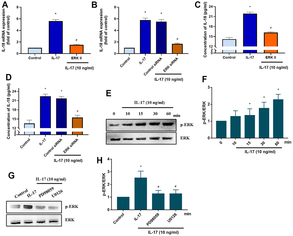
Figure 5.ERK signaling mediates IL-18 production in OASFs in response to IL-17 stimulation. (A) OASFs were pretreated with the ERK inhibitor ERKII (5 μM) for 1 h, then incubated with IL-17 (10 ng/mL) for 24 h. IL-18 expression was determined by qPCR. (B) OASFs were transfected with ERK siRNA or control siRNA for 24 h, then stimulated with IL-17 (10 ng/mL) for a further 24 h. IL-18 expression was assessed by qPCR. (C, D) OASFs were treated as described in Figure 5A, 5B. IL-18 production was examined by ELISA. (E, F) OASFs were stimulated with IL-17 (10 ng/mL) for different time intervals (0–60 min). The total cell lysates were collected and Western blot assessed ERK protein phosphorylation. The quantification of blot was shown in Figure 5F. (G, H) OASFs were pretreated with PD98059 (10 μM) or U0126 (5 μM) for 1 h, then stimulated with IL-17 (10 ng/mL) for 60 min. The total cell lysates were collected and Western blot assessed ERK protein phosphorylation. ERK protein was used as the internal control. The quantification of blot was shown in Figure 5H. Results are expressed as the means ± S.D. *p<0.05 compared with controls; #p<0.05 compared with the IL-17-treated group.
Figure 5 — IL-17 promotes IL-18 production via the MEK/ERK/miR-4492 axis in osteoarthritis synovial fibroblasts | Aging