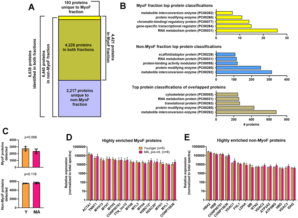
Figure 1.MyoF and non-MyoF protein characteristics. Legend: Data presented for MA (pre-intervention) and Y include the total number of proteins identified in each fraction (A), the top 5 protein classifications from each fraction (B), the number of MyoF and non-MyoF proteins detected within and between age cohorts (C), the top 15 highly enriched MyoF proteins (D), and the top 15 highly enriched non-MyoF proteins (E). Data in panels c and d are presented as means with standard deviation bars, and y-axes were scaled as log10 for improved visualization. Symbols: *, indicates multiple histone isoforms were congregated into these two targets based on sequence similarities. Protein names for gene symbols in panel c: ACTA1, Actin Alpha 1, Skeletal Muscle; TNNT1, Troponin T1; MYH13/7/2/8/4/1, myosin heavy chain isoforms 13/7/2/8/4/1; TTN_iso12, titin, isoform 12; MYL3/1/, myosin light chain isoforms 3/1; H2BC12, Histone H2B type 1-K/C/E/F/G/I/type F-S; H2AC20, Histone H2A type 2-A/C. Protein names for gene symbols in panel d: HBA2, Hemoglobin subunit alpha; HBB, Hemoglobin subunit beta; VDAC1, Voltage-dependent anion-selective channel protein 1; FHL1, Four and a half LIM domains protein 1; LDHA, L-lactate dehydrogenase A chain; MB, myoglobin; RTN2, Isoform RTN2-C of Reticulon-2; VDAC2, Voltage-dependent anion-selective channel protein 2; ATP2A2, Sarcoplasmic/endoplasmic reticulum calcium ATPase 2; ATP5MG, ATP synthase subunit g, mitochondrial; HBD, Hemoglobin subunit delta; VDAC3, Voltage-dependent anion-selective channel protein 3; IDH2, Isocitrate dehydrogenase [NADP], mitochondrial. Other note: CONP00761/15636 are non-annotated proteins found in both fractions.