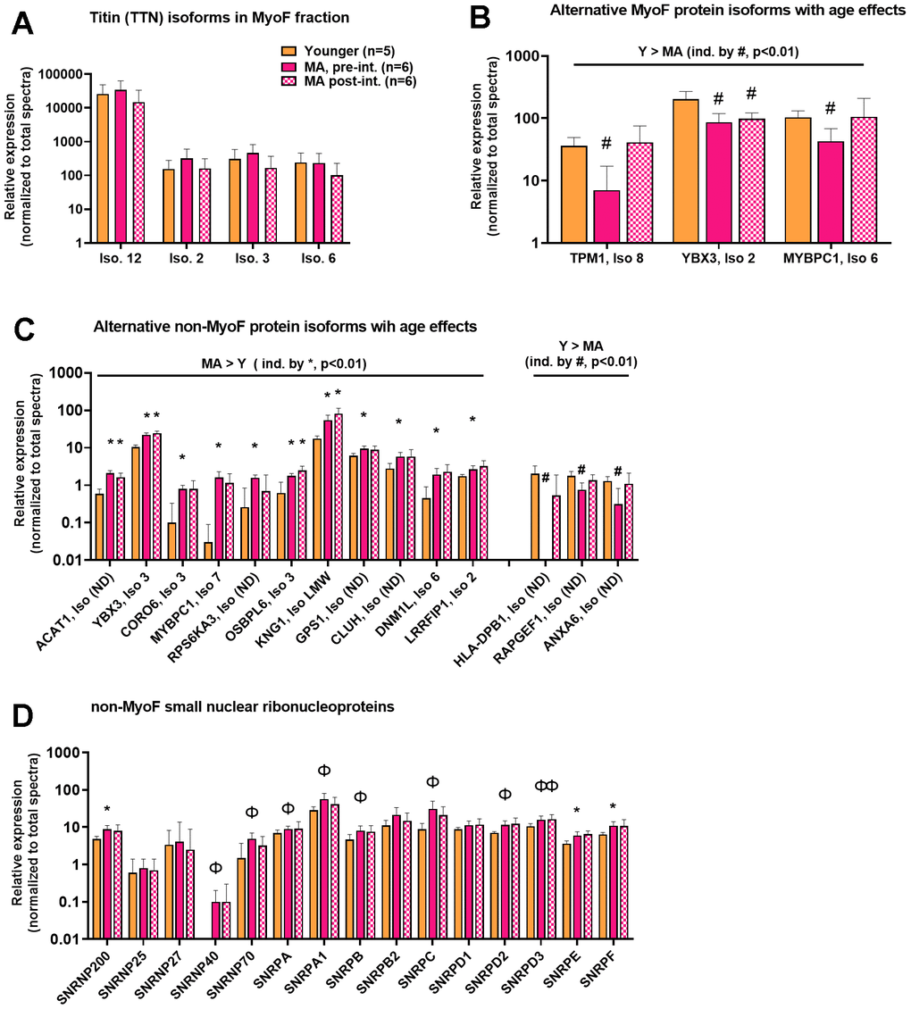
Figure 3.MyoF and non-MyoF alternative protein isoform differences detected with proteomics. Legend: Data presented for Y and MA (pre- and post-intervention) include the identified titin isoforms in the MyoF fraction (A), alternative MyoF protein isoforms affected by aging (B), and alternative non-MyoF protein isoforms affected by aging (C), and small nuclear ribonucleoproteins that make up spliceosomes between cohorts (D). Data are presented as mean ± standard deviations for individual protein spectra values (normalized to total run spectra values) and y-axes were scaled as log10 for improved visualization. Symbols: #, indicates lower in MA versus Y at one or both time points (p<0.01); *, indicates greater in MA versus Y at one or both time points (p<0.01); Φ, indicates greater in MA versus Y at one or both time points for panel d only (p<0.05). Notes: (ND), indicates that the isoform number was not provided from the Uniprot’s Homo Sapiens reference database (UP000005640_9606).