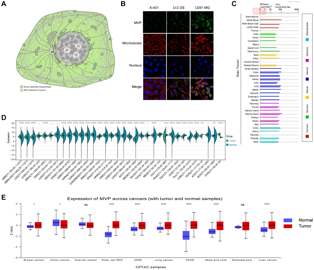
Figure 1.MVP localization, functional chaperone, and expression profiles in normal tissues and cancer. (A) MVP protein is localized in cytoplasm. (B) Immunofluorescence staining to detect the subcellular distribution of MVP in the nucleus, endoplasmic reticulum (ER) and microtubules of A-431 epidermoid carcinoma, U-2 osteosarcoma cells, and U251 glioblastoma. (C) Bar plot of MVP mRNA expression in various normal human tissues. (D) Expression levels of MVP mRNA in 34 different tumor types in the TCGA database via the SangerBox. (E) Expression levels of MVP protein in different tumors and corresponding normal tissues in the UALCAN portal. nsP ≥ 0.05, *P < 0.05, **P < 0.01, ***P < 0.001, ****P < 0.0001.