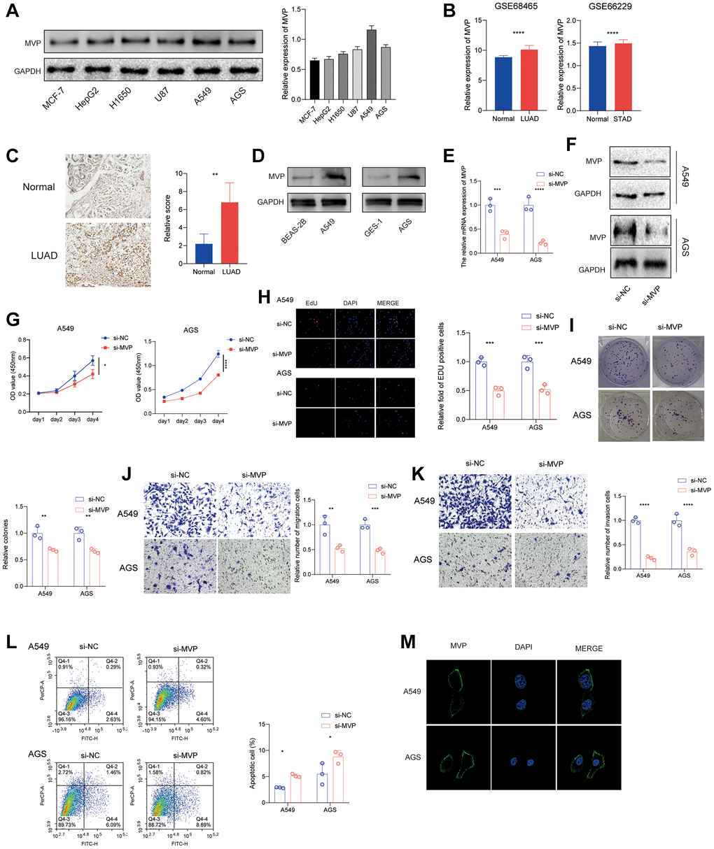
Figure 8.MVP knockdown effects in cancer cells. (A) The protein expression level of MVP in each cell line was tested by western blot. (B) Analysis of the GEO database showed that MVP was highly expressed in LUAD and STAD compared to adjacent tissues. (C) Immunohistochemical staining showed that MVP was highly expressed in LUAD compared with adjacent tissues. (D) Western blot assay confirmed that MVP expression was higher in A549 and AGS cells than in their corresponding normal epithelial cells. (E, F) RT-qPCR and western blot confirming MVP knockdown efficiency in A549 and AGS. (G) CCK-8 assay measuring cell proliferation in MVP-knockdown and control cells. (H) EdU assay measuring cell proliferation in MVP-knockdown and control cells. (I) Colony formation assay showed that MVP knockdown inhibits the colony formation ability of cells. (J, K) Transwell migration and invasion assays showing decreased migration and invasion of MVP-knockdown cells. (L) Cell apoptosis was monitored by flow cytometry. (M) Immunofluorescence assay showed that MVP was mainly distributed in the cell membrane and cytoplasm of A549 and AGS cells. *P < 0.05, **P < 0.01, ***P < 0.001, ****P < 0.0001.