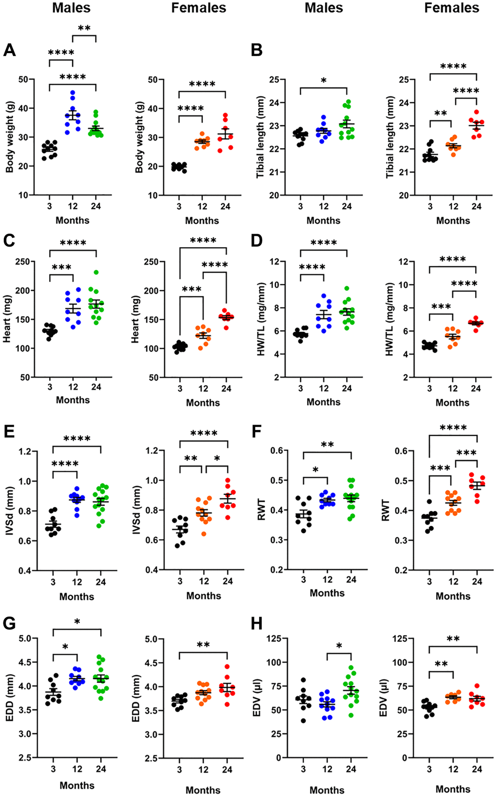
Figure 2.Effects of aging on the heart. For each panel, graphs of males (blue and green) and females (orange and red) are represented side by side. (A) Body weight, (B) Tibial length, (C) Heart weight, (D) indexed heart weight for tibial length (HW/TL), (E) Diastolic thickness of the interventricular septum (IVSd) by echo, (F) End-diastolic LV diameter (EDD), (G) Left ventricular relative wall thickness (RWT) and (H) End-diastolic LV volume (EDV). Data are represented as mean +SEM. One-way ANOVA followed by Holm-Sidak post-test. *p < 0.05, **p < 0.01, ***p < 0.001 and ****p < 0.0001 between indicated groups.