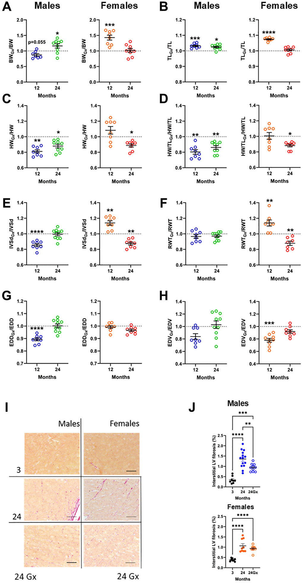
Figure 3.Loss of gonadal steroid hormones in young and adult mice and its effects on the heart later in life. (A–H) Results are illustrated as a ratio of individual values of indicated parameters in gonadectomized (Gx) animals on the mean value for this parameter from a group of age-matched controls. The dotted line indicates a ratio of 1 or the absence of difference between Gx and non-Gx animals. Blue and green (males) and orange and red (females) indicate the ratio in adult (12 months) and old mice (24 months). (A) Body weight, (B) Tibial length, (C) Heart weight, (D) indexed heart weight for tibial length (HW/TL), (E) Diastolic thickness of the interventricular septum (IVSd) by echo, (F) End-diastolic LV diameter (EDD), (G) Left ventricular relative wall thickness (RWT) and (H) End-diastolic LV volume (EDV). (I) Representative pictures of picrosirius-stained LV sections for the indicated groups. (J) Interstitial myocardial fibrosis in aging male and female mice, intact or Gx. Data are represented as mean +SEM. (A–H) Student T-test comparison with age-matched controls. (J) One-way ANOVA followed by Holm-Sidak post-test. *p < 0.05, **p < 0.01, ***p < 0.001 and ****p < 0.0001.