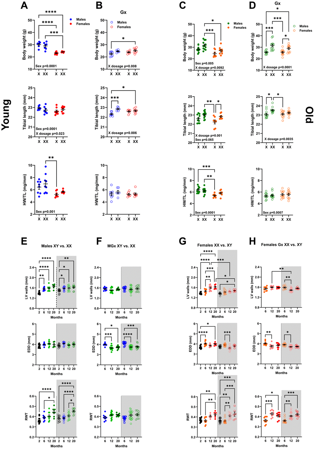
Figure 4.Sex chromosomes significantly affect body growth but little on cardiac growth and LV remodelling during aging. Body weight, Tibial length, and indexed Heart weight for tibial length (HW/TL). A and B: Young mice (6 months). (A) non-Gx 6-month-old mice with one (X) or two X chromosomes (XX). Phenotypic males (solid blue dots) and females (solid red dots). (B) Gx 6-month-old mice (Gx at five weeks). Phenotypic males (blue circles) and females (red circles). (C, D) Old mice (20 months). (C) non-Gx 20-month-old mice. Phenotypic males (solid green dots) and females (solid orange dots). (D) Gx 20-month-old mice (Gx at six months). Phenotypic males (green circles) and females (orange circles). Data are represented as mean +SEM. Two-way ANOVA followed by Holm-Sidak post-test. *p < 0.05, **p < 0.01, ***p < 0.001 and ****p < 0.0001 between indicated groups. Variables are phenotypical sex (Sex) and one or two X chromosomes (X dosage). P-values below 0.05 are indicated on the graphs for the two variables. (E–H) LV wall thickness, End-diastolic LV diameter (EDD) and LV relative wall thickness (RWT) during aging by echocardiography. (E) Non-Gx males with one X chromosome (left; solid dots) or two X chromosomes (right; empty dots and gray background). (F) Gx males (MGx) at the age of 6 months. (G) Non-Gx females with two X chromosomes (left; solid dots) or one X chromosome (right; empty dots and gray background). (H) Gx females (FGx) at the age of 6 months. Data are represented as mean +SEM. One-way ANOVA followed by Holm-Sidak post-test. *p < 0.05, **p < 0.01, ***p < 0.001 and ****p < 0.0001 between indicated groups.