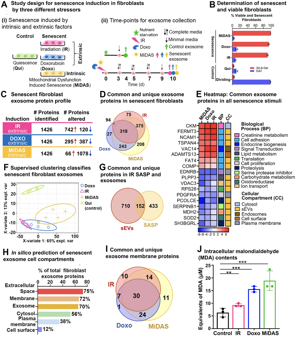
Figure 3.Senescence stimuli specific exosome protein signatures in primary human lung fibroblasts (IMR90). (A) (i). Study design showing three different senescence stimuli, Irradiation; IR, Doxorubicin; doxo, and Antimycin A-induced mitochondrial dysfunction-associated senescence; MiDAS, were used to generate senescent primary human lung fibroblasts (n=4, each condition), and Quiescent cells (Qui) were used as control. (ii) A timeline indicating treatment days and endpoints for the three different senescence inducers and control conditions. (B) Bar graph showing the percentage of viable and senescent fibroblasts in the different senescence-induction conditions using Edu (shown in blue) and SA-β-Gal (shown in red). (C) Summary of significantly altered exosome proteins (q-value < 0.05 and ≥ 1.5-fold change) in senescent human lung fibroblasts compared to quiescent cells following IR, doxo, and MiDAS senescence induction. (D) Venn diagram showing overlapping and unique protein signatures of senescent human lung fibroblast exosomes induced by IR, doxo, and MiDAS. (E) Heatmap displaying the exosome protein fold change averaged across replicates, showing the top 18 altered proteins, increased (red) and decreased (blue) due to senescence. The intensities averaged for Qui were used as the baseline. ClueGO biological pathways and cell compartments are presented. BP; Biological process, CC; Cellular compartment. (F) Partial least squares-discriminant analysis (PLS-DA) clustered senescent fibroblasts from Quiescent control. The two variates explaining the most significant variations are shown. (G) Venn diagram showing overlapping and unique exosomal SASP (sEVs) and soluble SASP protein signatures of IR-induced senescent fibroblasts. (H) Enrichment analysis of Gene Ontology/cellular compartments overrepresented among protein contents of senescent fibroblast exosomes. (I) Venn diagram showing overlapping and unique surface protein signatures of senescent fibroblast exosomes induced by IR, doxo, and MiDAS. (J) Bar graph showing malondialdehyde cellular content, a product of lipid peroxidation used as a marker for intracellular oxidative stress measured in the different senescence stimuli.