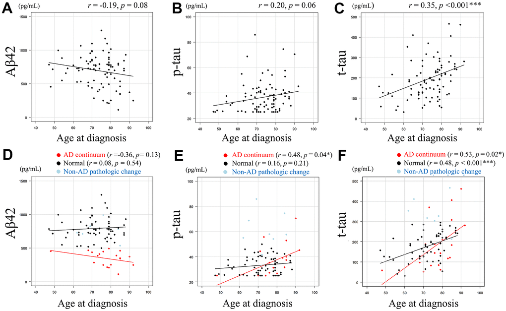
Figure 1.Relationship between Aβ42, p-tau, or t-tau levels and age at diagnosis. Scatter plots demonstrate the relationship between age at diagnosis and CSF biomarkers. Least squares regression lines are included where Pearson’s correlation analysis was performed. A negative correlative tendency (r = -0.19, p = 0.08) was observed between CSF Aβ42 levels (pg/mL) and age at diagnosis. (A) A positive correlative tendency (r = 0.20, p = 0.06) was observed between CSF p-tau levels (pg/mL) and age at diagnosis. (B) Significant positive correlation was observed between CSF t-tau levels (pg/mL) and age at diagnosis (r = 0.35, p < 0.001). (C) The association between age at diagnosis and each AT(N) category for CSF Aβ42, p-tau, and t-tau are shown. (D–F), with Pearson’s correlation coefficient not calculated for the non-AD category due to the small number of cases. CSF Aβ42 and age at diagnosis showed negative correlative tendency (r =-0.36, p= 0.13) in the AD continuum category, whereas this tendency was not evident in the normal category (r = 0.08, p = 0.54). (D) CSF p-tau and age at diagnosis showed a significant positive correlation in the AD continuum category (r = 0.48, p = 0.04), but showed no obvious tendency in the normal category. (E) CSF t-tau and age at diagnosis showed a significant positive correlation in both patients in AD continuum category (r = 0.53, p = 0.02) and normal category (r = 0.48, p < 0.001). (F) Data in AD continuum category are plotted in red, normal category in black, and non-AD pathologic change category in light blue (D–F). Aβ42 = amyloid-beta 42, p-tau = phosphorylated tau, t-tau = total tau, * = p < 0.05, *** = p < 0.001. r and p represent Pearson’s correlation and significance, respectively.