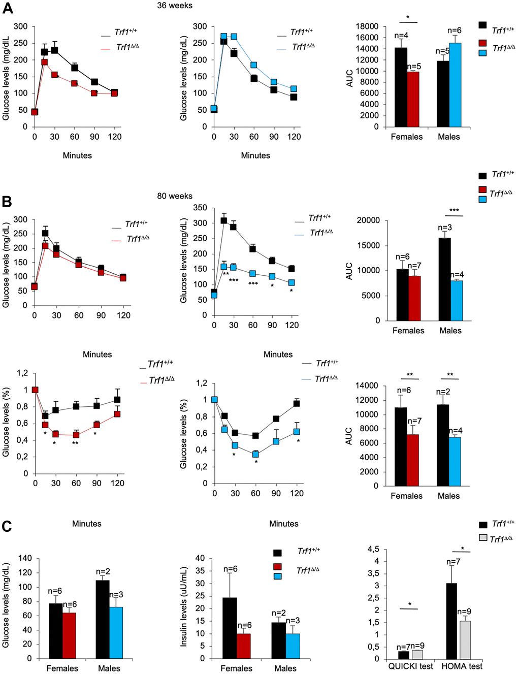
Figure 3.Trf1Δ/Δ mice show better tolerance to glucose. (A) Glucose tolerance test in 36-week-old-mice. Left: fasting glucose levels (mg/dl) measured at different time points (minutes) in females and males. Right: quantification of the area under the glucose tolerance test curve (AUC) in females and males. Note that Trf1 Δ/Δ females have lower glucose levels compared to wild-types. (B) Glucose and insulin tolerance test in 80-week-old-mice. Up: Glucose tolerance test. Left: fasting glucose levels (mg/dl) measured at different time points (minutes). Right: quantification of the area under the glucose tolerance test curve (AUC) in females and males. Bottom: Insulin tolerance test. Left: glucose levels (%) relative to glucose levels in fasting measured at different time points (minutes). Right: quantification of the area under the insulin tolerance test curve (AUC) in females and males. Note that Trf1Δ/Δ males have a better tolerance to glucose than wild-types. In addition, Trf1Δ/Δ mice showed better tolerance to insulin. (C) Fasting glucose and fasting insulin levels, derived HOMA-IR insulin-resistance quantification, and QUICKI insulin sensitivity quantification of 80-week-old males and females. Note that Trf1Δ/Δ have increased insulin sensitivity and improved insulin resistance shown by QUICKI and HOMA-IR index respectively. Error bars, s.e.m.; only significant values are shown; *P < 0.05; **P < 0.01; ***P < 0.001 determined by two-tailed Student’s t-test (A–C)